August 2025

Gout

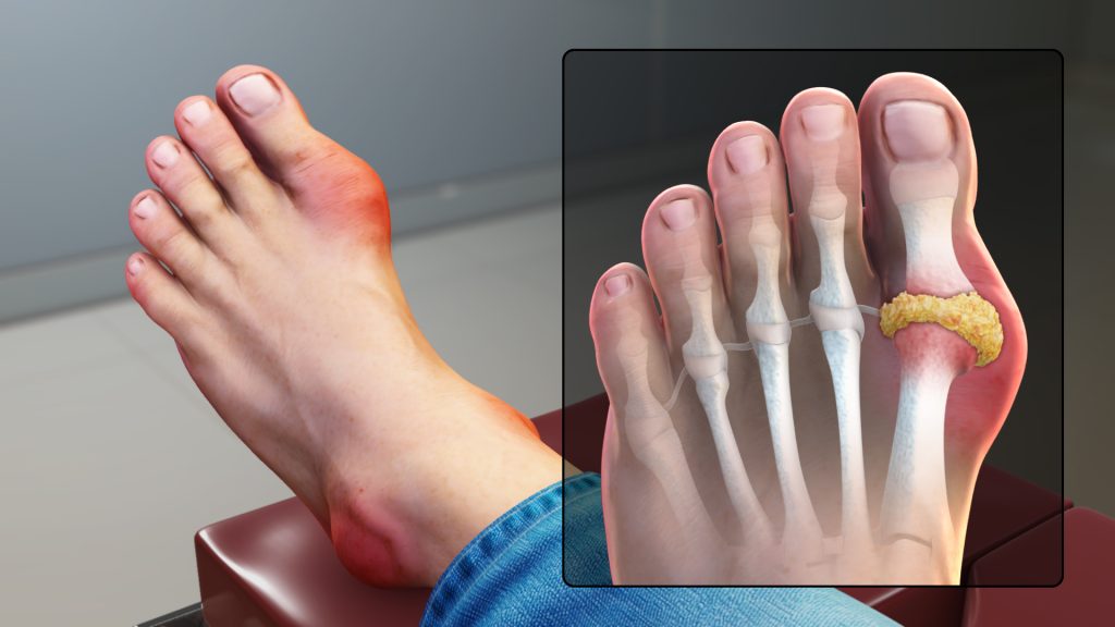
Gout Gout is a common and complex form of arthritis that can affect anyone. It’s characterized by sudden, severe attacks of pain, swelling, redness and tenderness in the joints, often the joint at the base of the big toe. An attack of gout can occur suddenly, often waking you up in the middle of the night with the sensation that your big toe is on fire. The affected joint is hot, swollen and so tender that even the weight of the sheet on it may seem intolerable. Gout symptoms may come and go, but there are ways to manage symptoms and prevent flares. Causes of Gout Gout occurs when urate crystals accumulate in your joint, causing the inflammation and intense pain of a gout attack. Urate crystals can form when you have high levels of uric acid in your blood. Your body produces uric acid when it breaks down purines substances that are found naturally in your body. Purines are also found in certain foods, such as steak, organ meats and seafood. Other foods also promote higher levels of uric acid, such as alcoholic beverages, especially beer, and drinks sweetened with fruit sugar (fructose). Normally, uric acid dissolves in your blood and passes through your kidneys into your urine. But sometimes either your body produces too much uric acid or your kidneys excrete too little uric acid. When this happens, uric acid can build up, forming sharp, needlelike urate crystals in a joint or surrounding tissue that cause pain, inflammation and swelling. Symptoms of Gout The signs and symptoms of gout almost always occur suddenly, and often at night. They include: Intense joint pain.  Gout usually affects the large joint of your big toe, but it can occur in any joint. Other commonly affected joints include the ankles, knees, elbows, wrists and fingers. The pain is likely to be most severe within the first four to 12 hours after it begins. Lingering discomfort.  After the most severe pain subsides, some joint discomfort may last from a few days to a few weeks. Later attacks are likely to last longer and affect more joints. Inflammation and redness.  The affected joint or joints become swollen, tender, warm and red. Limited range of motion.  As gout progresses, you may not be able to move your joints normally. When to see a doctor If you experience sudden, intense pain in a joint, call your doctor. Gout that goes untreated can lead to worsening pain and joint damage. Seek medical care immediately if you have a fever and a joint is hot and inflamed, which can be a sign of infection. Risk factors You’re more likely to develop gout if you have high levels of uric acid in your body. Factors that increase the uric acid level in your body include: Diet.  Eating a diet rich in meat and seafood and drinking beverages sweetened with fruit sugar (fructose) increase levels of uric acid, which increase your risk of gout. Alcohol consumption, especially of beer, also increases the risk of gout. Obesity.  If you’re overweight, your body produces more uric acid and your kidneys have a more difficult time eliminating uric acid. Medical conditions.  Certain diseases and conditions increase your risk of gout. These include untreated high blood pressure and chronic conditions such as diabetes, metabolic syndrome, and heart and kidney diseases. Certain medications.  The use of thiazide diuretics  commonly used to treat hypertension and low-dose aspirin also can increase uric acid levels. So can the use of anti-rejection drugs prescribed for people who have undergone an organ transplant. Family history of gout.  If other members of your family have had gout, you’re more likely to develop the disease. Age and sex.  Gout occurs more often in men, primarily because women tend to have lower uric acid levels. After menopause, however, women’s uric acid levels approach those of men. Men are also more likely to develop gout earlier usually between the ages of 30 and 50 whereas women generally develop signs and symptoms after menopause. Recent surgery or trauma.  Experiencing recent surgery or trauma has been associated with an increased risk of developing a gout attack. Complications People with gout can develop more-severe conditions, such as: Recurrent gout.  Some people may never experience gout signs and symptoms again. Others may experience gout several times each year. Medications may help prevent gout attacks in people with recurrent gout. If left untreated, gout can cause erosion and destruction of a joint. Advanced gout.  Untreated gout may cause deposits of urate crystals to form under the skin in nodules called tophi . Tophi can develop in several areas such as your fingers, hands, feet, elbows or Achilles tendons along the backs of your ankles. Tophi usually aren’t painful, but they can become swollen and tender during gout attacks. Kidney stones.  Urate crystals may collect in the urinary tract of people with gout, causing kidney stones. Medications can help reduce the risk of kidney stones. Prevention of Gout During symptom-free periods, these dietary guidelines may help protect against future gout attacks: Drink plenty of fluids.  Stay well-hydrated, including plenty of water. Limit how many sweetened beverages you drink, especially those sweetened with high-fructose corn syrup. Limit or avoid alcohol. Talk with your doctor about whether any amount or type of alcohol is safe for you. Recent evidence suggests that beer may be particularly likely to increase the risk of gout symptoms, especially in men. Get your protein from low-fat dairy products. Low-fat dairy products may actually have a protective effect against gout, so these are your best-bet protein sources. Limit your intake of meat, fish and poultry.  A small amount may be tolerable, but pay close attention to what types and how much seem to cause problems for you. Maintain a desirable body weight.  Choose portions that allow you to maintain a healthy weight. Losing weight may decrease uric acid levels in your body. But avoid fasting or rapid weight loss, since doing so may temporarily raise uric acid

Gout Read More »

Goiter

Goiter A goiter is an abnormal enlargement of your thyroid gland. Your thyroid is a butterfly-shaped gland located at the base of your neck just below your Adam’s apple. Although goiters are usually painless, a large goiter can cause a cough and make it difficult for you to swallow or breathe. The most common cause of goiters worldwide is a lack of iodine in the diet. In the United States, where the use of iodized salt is common, a goiter is more often due to the over- or underproduction of thyroid hormones or to nodules that develop in the gland itself. Treatment depends on the size of the goiter, your symptoms and the underlying cause. Small goiters that aren’t noticeable and don’t cause problems usually don’t need treatment. Causes of Goiter Your thyroid gland produces two main hormones  thyroxine (T-4) and triiodothyronine (T-3). These hormones circulate in your bloodstream and help regulate your metabolism. They maintain the rate at which your body uses fats and carbohydrates, help control your body temperature, influence your heart rate, and help regulate the production of proteins. Your thyroid gland also produces calcitonin a hormone that helps regulate the amount of calcium in your blood. Your pituitary gland and hypothalamus control the rate at which these hormones are produced and released. The process begins when the hypothalamus an area at the base of your brain that acts as a thermostat for your whole system signals your pituitary gland to make a hormone known as thyroid-stimulating hormone (TSH). Your pituitary gland also located at the base of your brain releases a certain amount of TSH, depending on how much thyroxine and T-3 are in your blood. Your thyroid gland, in turn, regulates its production of hormones based on the amount of TSH it receives from the pituitary gland. Having a goiter doesn’t necessarily mean that your thyroid gland isn’t working normally. Even when it’s enlarged, your thyroid may produce normal amounts of hormones. It might also, however, produce too much or too little thyroxine and T-3. A number of factors can cause your thyroid gland to enlarge. Among the most common are: Iodine deficiency. Iodine, which is essential for the production of thyroid hormones, is found primarily in seawater and in the soil in coastal areas. In the developing world, people who live inland or at high elevations are often iodine deficient and can develop goiters when the thyroid enlarges in an effort to obtain more iodine. The initial iodine deficiency may be made even worse by a diet high in hormone-inhibiting foods, such as cabbage, broccoli and cauliflower. Although a lack of dietary iodine is the main cause of goiters in many parts of the world, this is not often the case in countries where iodine is routinely added to table salt and other foods. Graves’ disease.  A goiter can sometimes occur when your thyroid gland produces too much thyroid hormone (hyperthyroidism). In Graves’ disease, antibodies produced by your immune system mistakenly attack your thyroid gland, causing it to produce excess thyroxine. This overstimulation causes the thyroid to swell. Hashimoto’s disease.  A goiter can also result from an underactive thyroid (hypothyroidism). Like Graves’ disease, Hashimoto’s disease is an autoimmune disorder. But instead of causing your thyroid to produce too much hormone, Hashimoto’s damages your thyroid so that it produces too little. Sensing a low hormone level, your pituitary gland produces more TSH to stimulate the thyroid, which then causes the gland to enlarge. Multinodular goiter.  In this condition, several solid or fluid-filled lumps called nodules develop in both sides of your thyroid, resulting in overall enlargement of the gland. Solitary thyroid nodules.  In this case, a single nodule develops in one part of your thyroid gland. Most nodules are noncancerous (benign) and don’t lead to cancer. Thyroid cancer.  Thyroid cancer is far less common than benign thyroid nodules. A biopsy of a thyroid nodule is very accurate in determining if it’s cancerous. Pregnancy.  A hormone produced during pregnancy, human chorionic gonadotropin (HCG), may cause your thyroid gland to enlarge slightly. Inflammation.  Thyroiditis is an inflammatory condition that can cause pain and swelling in the thyroid. It may also cause an over- or underproduction of thyroxine. Symptoms of Goiter Not all goiters cause signs and symptoms. When signs and symptoms do occur they may include: Risk factors Goiters can affect anyone. They may be present at birth and occur at any time throughout life. Some common risk factors for goiters include: A lack of dietary iodine.  People living in areas where iodine is in short supply and who don’t have access to iodine supplements are at high risk of goiters. Being female.  Because women are more prone to thyroid disorders, they’re also more likely to develop goiters. Your age.  Goiters are more common after age 40. Medical history.  A personal or family history of autoimmune disease increases your risk. Pregnancy and menopause.  For reasons that aren’t entirely clear, thyroid problems are more likely to occur during pregnancy and menopause. Certain medications.  Some medical treatments, including the heart drug amiodarone (Cordarone, Pacerone, others) and the psychiatric drug lithium (Lithobid, others), increase your risk. Radiation exposure.  Your risk increases if you’ve had radiation treatments to your neck or chest area or you’ve been exposed to radiation in a nuclear facility, test or accident. Complications Small goiters that don’t cause physical or cosmetic problems aren’t a concern. But large goiters can make it hard to breathe or swallow and can cause a cough and hoarseness. Goiters that result from other conditions, such as hypothyroidism or hyperthyroidism, can be associated with a number of symptoms, ranging from fatigue and weight gain to unintended weight loss, irritability and trouble sleeping. Diagnosis Your doctor may discover an enlarged thyroid gland simply by feeling your neck and having you swallow during a routine physical exam. In some cases, your doctor may also be able to feel the presence of nodules. Diagnosing a goiter may also involve: A hormone test.  Blood tests can

Goiter Read More »

Glaucoma

Glaucoma Glaucoma is a group of eye conditions that damage the optic nerve, the health of which is vital for good vision. This damage is often caused by an abnormally high pressure in your eye. Glaucoma is one of the leading causes of blindness for people over the age of 60. It can occur at any age but is more common in older adults. Many forms of glaucoma have no warning signs. The effect is so gradual that you may not notice a change in vision until the condition is at an advanced stage. Because vision loss due to glaucoma can’t be recovered, it’s important to have regular eye exams that include measurements of your eye pressure so a diagnosis can be made in its early stages and treated appropriately. If glaucoma is recognized early, vision loss can be slowed or prevented. If you have the condition, you’ll generally need treatment for the rest of your life. Causes of Glaucoma Glaucoma is the result of damage to the optic nerve. As this nerve gradually deteriorates, blind spots develop in your visual field. For reasons that doctors don’t fully understand, this nerve damage is usually related to increased pressure in the eye. Elevated eye pressure is due to a buildup of a fluid (aqueous humor) that flows throughout the inside of your eye. This internal fluid normally drains out through a tissue called the trabecular meshwork at the angle where the iris and cornea meet. When fluid is overproduced or the drainage system doesn’t work properly, the fluid can’t flow out at its normal rate and eye pressure increases. Glaucoma tends to run in families. In some people, scientists have identified genes related to high eye pressure and optic nerve damage. Types of glaucoma include: Open-angle glaucoma Open-angle glaucoma is the most common form of the disease. The drainage angle formed by the cornea and iris remains open, but the trabecular meshwork is partially blocked. This causes pressure in the eye to gradually increase. This pressure damages the optic nerve. It happens so slowly that you may lose vision before you’re even aware of a problem. Angle-closure glaucoma Angle-closure glaucoma, also called closed-angle glaucoma, occurs when the iris bulges forward to narrow or block the drainage angle formed by the cornea and iris. As a result, fluid can’t circulate through the eye and pressure increases. Some people have narrow drainage angles, putting them at increased risk of angle-closure glaucoma. Angle-closure glaucoma may occur suddenly (acute angle-closure glaucoma) or gradually (chronic angleclosure glaucoma). Acute angle-closure glaucoma is a medical emergency. Normal-tension glaucoma In normal-tension glaucoma, your optic nerve becomes damaged even though your eye pressure is within the normal range. No one knows the exact reason for this. You may have a sensitive optic nerve, or you may have less blood being supplied to your optic nerve. This limited blood flow could be caused by atherosclerosis the buildup of fatty deposits (plaque) in the arteries or other conditions that impair circulation. Glaucoma in children It’s possible for infants and children to have glaucoma. It may be present from birth or develop in the first few years of life. The optic nerve damage may be caused by drainage blockages or an underlying medical condition. Pigmentary glaucoma In pigmentary glaucoma, pigment granules from your iris build up in the drainage channels, slowing or blocking fluid exiting your eye. Activities such as jogging sometimes stir up the pigment granules, depositing them on the trabecular meshwork and causing intermittent pressure elevations. Symptoms of Glaucoma The signs and symptoms of glaucoma vary depending on the type and stage of your condition. For example: Open-angle glaucoma Acute angle-closure glaucoma N.B – If left untreated, glaucoma will eventually cause blindness. Even with treatment, about 15 percent of people with glaucoma become blind in at least one eye within 20 years. When to see a doctor Promptly go to an emergency room or an eye doctor’s (ophthalmologist’s) office if you experience some of the symptoms of acute angle-closure glaucoma, such as severe headache, eye pain and blurred vision. Risk factors Because chronic forms of glaucoma can destroy vision before any signs or symptoms are apparent, be aware of these risk factors: Prevention These self-care steps can help you detect glaucoma in its early stages, which is important in preventing vision loss or slowing its progress. Get regular dilated eye examinations.  Regular comprehensive eye exams can help detect glaucoma in its early stages, before significant damage occurs. As a general rule, the American Academy of Ophthalmology recommends having a comprehensive eye exam every five to 10 years if you’re under 40 years old, every two to four years if you’re 40 to 54 years old; every one to three years if you’re 55 to 64 years old, and every one to two years if you’re older than 65. If you’re at risk of glaucoma, you’ll need more frequent screening. Ask your doctor to recommend the right screening schedule for you. Know your family’s eye health history.  Glaucoma tends to run in families. If you’re at increased risk, you may need more frequent screening. Exercise safely.  Regular, moderate exercise may help prevent glaucoma by reducing eye pressure. Talk with your doctor about an appropriate exercise program. Take prescribed eyedrops regularly.  Glaucoma eyedrops can significantly reduce the risk that high eye pressure will progress to glaucoma. To be effective, eyedrops prescribed by your doctor need to be used regularly even if you have no symptoms. Wear eye protection.  Serious eye injuries can lead to glaucoma. Wear eye protection when using power tools or playing highspeed racket sports in enclosed courts. Diagnosis Your doctor will review your medical history and conduct a comprehensive eye examination. He or she may perform several tests, including: Treatment The damage caused by glaucoma can’t be reversed. But treatment and regular checkups can help slow or prevent vision loss, especially if you catch the disease in its early stages. Glaucoma is treated by lowering your eye pressure (intraocular

Glaucoma Read More »

Ear infection (middle ear)

Ear infection (middle ear) An ear infection (sometimes called acute otitis media) is an infection of the middle ear, the air-filled space behind the eardrum that contains the tiny vibrating bones of the ear. Children are more likely than adults to get ear infections. Because ear infections often clear up on their own, treatment may begin with managing pain and monitoring the problem. Sometimes, antibiotics are used to clear the infection. Some people are prone to having multiple ear infections. This can cause hearing problems and other serious complications. Causes of Ear infection An ear infection is caused by a bacterium or virus in the middle ear. This infection often results from another illness cold, flu or allergy that causes congestion and swelling of the nasal passages, throat and eustachian tubes. Role of eustachian tubes The eustachian tubes are a pair of narrow tubes that run from each middle ear to high in the back of the throat, behind the nasal passages. The throat end of the tubes open and close to: N.B – Swollen eustachian tubes can become blocked, causing fluids to build up in the middle ear. This fluid can become infected and cause the symptoms of an ear infection. In children, the eustachian tubes are narrower and more horizontal, which makes them more difficult to drain and more likely to get clogged. Role of adenoids Adenoids are two small pads of tissues high in the back of the nose believed to play a role in immune system activity. Because adenoids are near the opening of the eustachian tubes, swelling of the adenoids may block the tubes. This can lead to middle ear infection. Swelling and irritation of adenoids is more likely to play a role in ear infections in children because children have relatively larger adenoids compared to adults. Related conditions Conditions of the middle ear that may be related to an ear infection or result in similar middle ear problems include: Otitis media with effusion, or swelling and fluid buildup (effusion) in the middle ear without bacterial or viral infection. This may occur because the fluid buildup persists after an ear infection has gotten better. It may also occur because of some dysfunction or noninfectious blockage of the eustachian tubes. Chronic otitis media with effusion, occurs when fluid remains in the middle ear and continues to return without bacterial or viral infection. This makes children susceptible to new ear infections and may affect hearing. Chronic suppurative otitis media, an ear infection that doesn’t go away with the usual treatments. This can lead to a hole in the eardrum. Symptoms of Ear infection The onset of signs and symptoms of ear infection is usually rapid. Children Signs and symptoms common in children include: Adults Common signs and symptoms in adults include: When to see a doctor Signs and symptoms of an ear infection can indicate a number of conditions. It’s important to get an accurate diagnosis and prompt treatment. Call your child’s doctor if: Risk factors Risk factors for ear infections include: Age Children between the ages of 6 months and 2 years are more susceptible to ear infections because of the size and shape of their eustachian tubes and because their immune systems are still developing. Group child care.  Children cared for in group settings are more likely to get colds and ear infections than are children who stay home. The children in group settings are exposed to more infections, such as the common cold. Infant feeding.  Babies who drink from a bottle, especially while lying down, tend to have more ear infections than do babies who are breast-fed. Seasonal factors.  Ear infections are most common during the fall and winter. People with seasonal allergies may have a greater risk of ear infections when pollen counts are high. Poor air quality.  Exposure to tobacco smoke or high levels of air pollution can increase the risk of ear infections. Cleft palate.  Differences in the bone structure and muscles in children who have cleft palates may make it more difficult for the eustachian tube to drain. Complications Most ear infections don’t cause long-term complications. Ear infections that happen again and again can lead to serious complications: Impaired hearing.  Mild hearing loss that comes and goes is fairly common with an ear infection, but it usually gets better after the infection clears. Ear infections that happen again and again, or fluid in the middle ear, may lead to more-significant hearing loss. If there is some permanent damage to the eardrum or other middle ear structures, permanent hearing loss may occur. Speech or developmental delays.  If hearing is temporarily or permanently impaired in infants and toddlers, they may experience delays in speech, social and developmental skills. Spread of infection.  Untreated infections or infections that don’t respond well to treatment can spread to nearby tissues. Infection of the mastoid, the bony protrusion behind the ear, is called mastoiditis. This infection can result in damage to the bone and the formation of pus-filled cysts. Rarely, serious middle ear infections spread to other tissues in the skull, including the brain or the membranes surrounding the brain (meningitis). Tearing of the eardrum.  Most eardrum tears heal within 72 hours. In some cases, surgical repair is needed. Prevention The following tips may reduce the risk of developing ear infections: Prevent common colds and other illnesses.  Teach your children to wash their hands frequently and thoroughly and to not share eating and drinking utensils. Teach your children to cough or sneeze into the crook of their arm. If possible, limit the time your child spends in group child care. A child care setting with fewer children may help. Try to keep your child home from child care or school when ill. Avoid secondhand smoke.  Make sure that no one smokes in your home. Away from home, stay in smoke-free environments. Breast-feed your baby.  If possible, breast-feed your baby for at least six months. Breast milk contains antibodies that may offer protection from ear infections. If

Ear infection (middle ear) Read More »

Dyslexia

Dyslexia Dyslexia is a learning disorder that involves difficulty reading due to problems identifying speech sounds and learning how they relate to letters and words (decoding). Also called reading disability, dyslexia affects areas of the brain that process language. People with dyslexia have normal intelligence and usually have normal vision. Most children with dyslexia can succeed in school with tutoring or a specialized education program. Emotional support also plays an important role. Though there’s no cure for dyslexia, early assessment and intervention result in the best outcome. Sometimes dyslexia goes undiagnosed for years and isn’t recognized until adulthood, but it’s never too late to seek help. Causes of Dyslexia Dyslexia tends to run in families. It appears to be linked to certain genes that affect how the brain processes reading and language, as well as risk factors in the environment. Symptoms of Dyslexia Signs of dyslexia can be difficult to recognize before your child enters school, but some early clues may indicate a problem. Once your child reaches school age, your child’s teacher may be the first to notice a problem. Severity varies, but the condition often becomes apparent as a child starts learning to read. Signs that a young child may be at risk of dyslexia include: Once your child is in school, dyslexia signs and symptoms may become more apparent, including: Teens and adults Dyslexia signs in teens and adults are similar to those in children. Some common dyslexia signs and symptoms in teens and adults include: When to see a doctor Though most children are ready to learn reading by kindergarten or first grade, children with dyslexia often can’t grasp the basics of reading by that time. Talk with your doctor if your child’s reading level is below what’s expected for his or her age or if you notice other signs of dyslexia. When dyslexia goes undiagnosed and untreated, childhood reading difficulties continue into adulthood. Risk factors Dyslexia risk factors include: Complications Dyslexia can lead to a number of problems, including: Trouble learning.  Because reading is a skill basic to most other school subjects, a child with dyslexia is at a disadvantage in most classes and may have trouble keeping up with peers. Social problems.  Left untreated, dyslexia may lead to low self-esteem, behavior problems, anxiety, aggression, and withdrawal from friends, parents and teachers. Problems as adults.  The inability to read and comprehend can prevent a child from reaching his or her potential as the child grows up. This can have long-term educational, social and economic consequences. Children who have dyslexia are at increased risk of having attention-deficit/hyperactivity disorder (ADHD), and vice versa. ADHD can cause difficulty sustaining attention as well as hyperactivity and impulsive behavior, which can make dyslexia harder to treat. Diagnosis There’s no single test that can diagnose dyslexia. A number of factors are considered, such as: Your child’s development, educational issues and medical history.  The doctor will likely ask you questions about these areas and want to know about any conditions that run in the family, including whether any family members have a learning disability. Home life.  The doctor may ask for a description of your family and home life, including who lives at home and whether there are any problems at home. Questionnaires.  The doctor may have your child, family members or teachers answer written questions. Your child may be asked to take tests to identify reading and language abilities. Vision, hearing and brain (neurological) tests.  These can help determine whether another disorder may be causing or adding to your child’s poor reading ability. Psychological testing.  The doctor may ask you and your child questions to better understand your child’s mental health. This can help determine whether social problems, anxiety or depression may be limiting your child’s abilities. Testing reading and other academic skills.  Your child may take a set of educational tests and have the process and quality of reading skills analyzed by a reading expert. Treatment There’s no known way to correct the underlying brain abnormality that causes dyslexia dyslexia is a lifelong problem. However, early detection and evaluation to determine specific needs and appropriate treatment can improve success. Educational techniques Dyslexia is treated using specific educational approaches and techniques, and the sooner the intervention begins, the better. Psychological testing will help your child’s teachers develop a suitable teaching program. Teachers may use techniques involving hearing, vision and touch to improve reading skills. Helping a child use several senses to learn for example, listening to a taped lesson and tracing with a finger the shape of the letters used and the words spoken can help in processing the information. Treatment focuses on helping your child: Individual education plan In the Zimbabwe, schools have a legal obligation to take steps to help children diagnosed with dyslexia with their learning problems. Talk to your child’s teacher about setting up a meeting to create a structured, written plan that outlines your child’s needs and how the school will help him or her succeed. This is called an Individualized Education Plan (IEP). Early treatment Children with dyslexia who get extra help in kindergarten or first grade often improve their reading skills enough to succeed in grade school and high school. Children who don’t get help until later grades may have more difficulty learning the skills needed to read well. They’re likely to lag behind academically and may never be able to catch up. A child with severe dyslexia may never have an easy time reading, but he or she can learn skills that improve reading and develop strategies to improve school performance and quality of life. What parents can do? You play a key role in helping your child succeed. Take these steps: Address the problem early.  If you suspect your child has dyslexia, talk to your child’s doctor. Early intervention can improve success. Read aloud to your child.  It’s best if you start when your child is 6 months old or even younger. Try listening to recorded books with your child. When

Dyslexia Read More »

Diaper rash

Diaper rash Diaper rash is a common form of inflamed skin (dermatitis) that appears as a patchwork of bright red skin on your baby’s bottom. Diaper rash is often related to wet or infrequently changed diapers, skin sensitivity, and chafing. It usually affects babies, though anyone who wears a diaper regularly can develop the condition. Diaper rash can alarm parents and annoy babies. But it usually clears up with simple at-home treatments, such as air drying, more frequent diaper changes and ointment. Causes of Diaper rash Diaper rash can be traced to a number of sources, including: Irritation from stool and urine.  Prolonged exposure to urine or stool can irritate a baby’s sensitive skin. Your baby may be more prone to diaper rash if he or she is experiencing frequent bowel movements or diarrhea because feces are more irritating than urine. Chafing or rubbing.  Tightfitting diapers or clothing that rubs against the skin can lead to a rash. Irritation from a new product.  Your baby’s skin may react to baby wipes, a new brand of disposable diapers, or a detergent, bleach or fabric softener used to launder cloth diapers. Other substances that can add to the problem include ingredients found in some baby lotions, powders and oils. Bacterial or yeast (fungal) infection.  What begins as a simple skin infection may spread to the surrounding region. The area covered by a diaper buttocks, thighs and genitals is especially vulnerable because it’s warm and moist, making a perfect breeding ground for bacteria and yeast. These rashes can be found within the creases of the skin, and there may be red dots scattered around the creases. Introduction of new foods. As babies start to eat solid foods, the content of their stool changes. This increases the likelihood of diaper rash. Changes in your baby’s diet can also increase the frequency of stools, which can lead to diaper rash. If your baby is breast-fed, he or she may develop diaper rash in response to something the mother has eaten. Sensitive skin.  Babies with skin conditions, such as atopic dermatitis or seborrheic dermatitis (eczema), may be more likely to develop diaper rash. However, the irritated skin of atopic dermatitis and eczema primarily affects areas other than the diaper area. Use of antibiotics.  Antibiotics kill bacteria the good kinds as well as the bad. When a baby takes antibiotics, bacteria that keep yeast growth in check may be depleted, resulting in diaper rash due to yeast infection. Antibiotic use also increases the risk of diarrhea. Breast-fed babies whose mothers take antibiotics are also at increased risk of diaper rash. Symptoms of Diaper rash Diaper rash is characterized by the following: Skin signs.  Diaper rash is marked by red, tender-looking skin in the diaper region buttocks, thighs and genitals. Changes in your baby’s disposition.  You may notice your baby seems more uncomfortable than usual, especially during diaper changes. A baby with a diaper rash often fusses or cries when the diaper area is washed or touched. When to see a doctor If your baby’s skin doesn’t improve after a few days of home treatment, talk with your doctor. Sometimes, you’ll need a prescription medication to treat diaper rash. Have your child examined if the rash: Prevention The best way to prevent diaper rash is to keep the diaper area clean and dry. A few simple strategies can help decrease the likelihood of diaper rash developing on your baby’s skin. Change diapers often.  Remove wet or dirty diapers promptly. If your child is in child care, ask staff members to do the same. Rinse your baby’s bottom with warm water as part of each diaper change.  You can use a sink, tub or water bottle for this purpose. Moist washcloths, cotton balls and baby wipes can aid in cleaning the skin, but be gentle. Don’t use wipes with alcohol or fragrance. If you wish to use soap, select a mild, fragrance-free type. Gently pat the skin dry with a clean towel or let it air dry.  Don’t scrub your baby’s bottom. Scrubbing can further irritate the skin. Don’t overtighten diapers.  Tight diapers prevent airflow into the diaper region, which sets up a moist environment favorable to diaper rashes. Tight diapers can also cause chafing at the waist or thighs. Give your baby’s bottom more time without a diaper.  When possible, let your baby go without a diaper. Exposing skin to air is a natural and gentle way to let it dry. To avoid messy accidents, try laying your baby on a large towel and engage in some playtime while he or she is bare-bottomed. Consider using ointment regularly.  If your baby gets rashes often, apply a barrier ointment during each diaper change to prevent skin irritation. Petroleum jelly and zinc oxide are the time-proven ingredients in many diaper ointments. After changing diapers, wash your hands well.  Hand-washing can prevent the spread of bacteria or yeast to other parts of your baby’s body, to you or to other children. In the past, it was common to use powders, such as cornstarch or talcum powder, to protect a baby’s skin and absorb excess moisture. Doctors no longer recommend this. Inhaled powder can irritate a baby’s lungs. Cloth or disposable diapers? Many parents wonder about what kind of diapers to use.  When it comes to preventing diaper rash, there’s no compelling evidence that cloth diapers are better than disposable diapers or vice versa. Because there’s no one best diaper, use whatever works for you and your baby.  If one brand of disposable diaper irritates your baby’s skin, try another. If the laundry soap you use on cloth diapers seems to cause a diaper rash, switch products. Whether you use cloth diapers, disposables or both kinds, always change your baby as soon as possible after he or she wets or soils the diaper to keep the bottom as clean and dry as possible. Washing cloth diapers If you use cloth diapers, careful washing can help prevent diaper rash. Washing methods vary

Diaper rash Read More »

Dementia

Dementia Dementia describes a group of symptoms affecting memory, thinking and social abilities severely enough to interfere with your daily life. It isn’t a specific disease, but several different diseases may cause dementia. Though dementia generally involves memory loss, memory loss has different causes. Having memory loss alone doesn’t mean you have dementia. Alzheimer’s disease is the most common cause of a progressive dementia in older adults, but there are a number of causes of dementia. Depending on the cause, some dementia symptoms may be reversible. Causes of Dementia Dementia is caused by damage to or loss of nerve cells and their connections in the brain. Depending on the area of the brain that’s affected by the damage, dementia can affect people differently and cause different symptoms. Dementias are often grouped by what they have in common, such as the protein or proteins deposited in the brain or the part of the brain that’s affected. Some diseases look like dementias, such as those caused by a reaction to medications or vitamin deficiencies, and they might improve with treatment. Progressive dementias Types of dementias that progress and aren’t reversible include: Alzheimer’s disease. Alzheimer’s disease is the most common cause of dementia. Although not all causes of Alzheimer’s disease are known, experts do know that a small percentage are related to mutations of three genes, which can be passed down from parent to child. While several different genes are probably involved in Alzheimer’s disease, one important gene that increases risk is apolipoprotein E4 (APOE). Alzheimer’s disease patients have plaques and tangles in their brains.  Plaques are clumps of a protein called beta-amyloid, and tangles are fibrous tangles made up of tau protein. It’s thought that these clumps damage healthy neurons and the fibers connecting them. Other genetic factors might make it more likely that people will develop Alzheimer’s. Vascular dementia.  This second most common type of dementia is caused by damage to the vessels that supply blood to your brain. Blood vessel problems can cause strokes or damage the brain in other ways, such as by damaging the fibers in the white matter of the brain. The most common symptoms of vascular dementia include difficulties with problem-solving, slowed thinking, focus and organization. These tend to be more noticeable than memory loss. Lewy body dementia.  Lewy bodies are abnormal balloonlike clumps of protein that have been found in the brains of people with Lewy body dementia, Alzheimer’s disease and Parkinson’s disease. This is one of the more common types of progressive dementia. Common signs and symptoms include acting out one’s dreams in sleep, seeing things that aren’t there (visual hallucinations), and problems with focus and attention. Other signs include uncoordinated or slow movement, tremors, and rigidity (parkinsonism). Frontotemporal dementia.  This is a group of diseases characterized by the breakdown (degeneration) of nerve cells and their connections in the frontal and temporal lobes of the brain, the areas generally associated with personality, behavior and language. Common symptoms affect behavior, personality, thinking, judgment, and language and movement. Mixed dementia.  Autopsy studies of the brains of people 80 and older who had dementia indicate that many had a combination of several causes, such as Alzheimer’s disease, vascular dementia and Lewy body dementia. Studies are ongoing to determine how having mixed dementia affects symptoms and treatments. Other disorders linked to dementia Huntington’s disease. Caused by a genetic mutation, this disease causes certain nerve cells in your brain and spinal cord to waste away. Signs and symptoms, including a severe decline in thinking (cognitive) skills, usually appear around age 30 or 40. Traumatic brain injury (TBI).  This condition is most often caused by repetitive head trauma. People such as boxers, football players or soldiers might experience TBI. Depending on the part of the brain that’s injured, this condition can cause dementia signs and symptoms such as depression, explosiveness, memory loss and impaired speech. TBI may also cause parkinsonism. Symptoms might not appear until years after the trauma. Creutzfeldt-Jakob disease.  This rare brain disorder usually occurs in people without known risk factors. This condition might be due to deposits of infectious proteins called prions. Creutzfeldt-Jakob disease usually has no known cause but can be inherited. It may also be caused by exposure to diseased brain or nervous system tissue, such as from a cornea transplant. Signs and symptoms of this fatal condition usually appear after age 60. Parkinson’s disease.  Many people with Parkinson’s disease eventually develop dementia symptoms (Parkinson’s disease dementia). Dementia-like conditions that can be reversed Some causes of dementia or dementia-like symptoms can be reversed with treatment. They include: Infections and immune disorders.  Dementia-like symptoms can result from fever or other side effects of your body’s attempt to fight off an infection. Multiple sclerosis and other conditions caused by the body’s immune system attacking nerve cells also can cause dementia. Metabolic problems and endocrine abnormalities.  People with thyroid problems, low blood sugar (hypoglycemia), too little or too much sodium or calcium, or problems absorbing vitamin B-12 can develop dementia-like symptoms or other personality changes. Nutritional deficiencies.   Not drinking enough liquids (dehydration); not getting enough thiamin (vitamin B-1), which is common in people with chronic alcoholism; and not getting enough vitamins B-6 and B-12 in your diet can cause dementia-like symptoms. Copper and vitamin E deficiencies also can cause dementia symptoms. Medication side effects.  Side effects of medications, a reaction to a medication or an interaction of several medications can cause dementia-like symptoms. Subdural hematomas.  Bleeding between the surface of the brain and the covering over the brain, which is common in the elderly after a fall, can cause symptoms similar to those of dementia. Poisoning.  Exposure to heavy metals, such as lead, and other poisons, such as pesticides, as well as recreational drug or heavy alcohol use can lead to symptoms of dementia. Symptoms might resolve with treatment. Brain tumors.  Rarely, dementia can result from damage caused by a brain tumor. Anoxia.  This condition, also called hypoxia, occurs when organ tissues aren’t getting enough oxygen.

Dementia Read More »

Cerebral palsy

Cerebral palsy Cerebral palsy is a group of disorders that affect movement and muscle tone or posture. It’s caused by damage that occurs to the immature brain as it develops, most often before birth. Signs and symptoms appear during infancy or preschool years. In general, cerebral palsy causes impaired movement associated with abnormal reflexes, floppiness or rigidity of the limbs and trunk, abnormal posture, involuntary movements, unsteady walking, or some combination of these. People with cerebral palsy can have problems swallowing and commonly have eye muscle imbalance, in which the eyes don’t focus on the same object. They also might have reduced range of motion at various joints of their bodies due to muscle stiffness. Cerebral palsy’s effect on function varies greatly. Some affected people can walk; others need assistance. Some people show normal or near-normal intellect, but others have intellectual disabilities. Epilepsy, blindness or deafness also might be present. Causes Cerebral palsy Cerebral palsy is caused by an abnormality or disruption in brain development, most often before a child is born. In many cases, the cause isn’t known. Factors that can lead to problems with brain development include: Symptoms Cerebral palsy Signs and symptoms can vary greatly. Movement and coordination problems associated with cerebral palsy include: N.B – Cerebral palsy can affect the whole body, or it might be limited primarily to one limb or one side of the body. The brain disorder causing cerebral palsy doesn’t change with time, so the symptoms usually don’t worsen with age. However, as the child gets older, some symptoms might become more or less apparent. And muscle shortening and muscle rigidity can worsen if not treated aggressively. Brain abnormalities associated with cerebral palsy might also contribute to other neurological problems, including: When to see a doctor It’s important to get a prompt diagnosis for a movement disorder or delays in your child’s development. See your child’s doctor if you have concerns about episodes of loss of awareness of surroundings or of abnormal bodily movements, abnormal muscle tone, impaired coordination, swallowing difficulties, eye muscle imbalance or other developmental issues. Risk factors A number of factors are associated with an increased risk of cerebral palsy. Maternal health Certain infections or toxic exposures during pregnancy can significantly increase cerebral palsy risk to the baby. Infections of particular concern include: Cytomegalovirus.  This common virus causes flu-like symptoms and can lead to birth defects if a mother has her first active infection during pregnancy. German measles (rubella).  This viral infection can be prevented with a vaccine. Herpes.  This can be passed from mother to child during pregnancy, affecting the womb and placenta. Inflammation triggered by infection can damage the unborn baby’s developing nervous system. Syphilis.  This is a sexually transmitted bacterial infection. Toxoplasmosis.  This infection is caused by a parasite found in contaminated food, soil and the feces of infected cats. Zika virus infection.  Infants for whom maternal Zika infection causes their head size to be smaller than normal (microcephaly) can develop cerebral palsy. Other conditions.  Other conditions that can increase the risk of cerebral palsy include thyroid problems, intellectual disabilities or seizures, and exposure to toxins, such as methyl mercury. Infant illness Illnesses in a newborn baby that can greatly increase the risk of cerebral palsy include: Bacterial meningitis.  This bacterial infection causes inflammation in the membranes surrounding the brain and spinal cord. Viral encephalitis.  This viral infection similarly causes inflammation in the membranes surrounding the brain and spinal cord. Severe or untreated jaundice.  Jaundice appears as a yellowing of the skin. The condition occurs when certain byproducts of “used” blood cells aren’t filtered from the bloodstream. Bleeding into the brain.  This condition is commonly caused by the baby having a stroke in the womb. Other factors of pregnancy and birth While the potential contribution from each is limited, additional pregnancy or birth factors associated with increased cerebral palsy risk include: Breech presentation.  Babies with cerebral palsy are more likely to be in this feet-first position at the beginning of labor rather than being headfirst. Low birth weight.  Babies who weigh less than 5.5 pounds (2.5 kilograms) are at higher risk of developing cerebral palsy. This risk increases as birth weight drops. Multiple babies.  Cerebral palsy risk increases with the number of babies sharing the uterus. If one or more of the babies die, the survivors’ risk of cerebral palsy increases. Premature birth.  Babies born fewer than 28 weeks into the pregnancy are at higher risk of cerebral palsy. The earlier a baby is born, the greater the cerebral palsy risk. Complications Muscle weakness, muscle spasticity and coordination problems can contribute to a number of complications either during childhood or in adulthood, including: Contracture.  Contracture is muscle tissue shortening due to severe muscle tightening (spasticity). Contracture can inhibit bone growth, cause bones to bend, and result in joint deformities, dislocation or partial dislocation. Premature aging.  Some type of premature aging will affect most people with cerebral palsy in their 40s because of the strain the condition puts on their bodies. Malnutrition.  Swallowing or feeding problems can make it difficult for someone who has cerebral palsy, particularly an infant, to get enough nutrition. This can impair growth and weaken bones. Some children need a feeding tube to get enough nutrition. Mental health conditions.  People with cerebral palsy might have mental health conditions, such as depression. Social isolation and the challenges of coping with disabilities can contribute to depression. Heart and lung disease.  People with cerebral palsy may develop heart disease and lung disease and breathing disorders. Osteoarthritis.  Pressure on joints or abnormal alignment of joints from muscle spasticity may lead to the early onset of this painful degenerative bone disease. Osteopenia.  Fractures due to low bone density (osteopenia) can stem from several common factors such as lack of mobility, nutritional shortcomings and anti-epileptic drug use. Prevention Most cases of cerebral palsy can’t be prevented, but you can lessen risks. If you’re pregnant or planning to become pregnant, you can take these steps to keep healthy and

Cerebral palsy Read More »

Acute liver failure

Acute liver failure Acute liver failure is loss of liver function that occurs rapidly  in days or weeks  usually in a person who has no pre-existing liver disease. Acute liver failure is less common than chronic liver failure, which develops more slowly. Acute liver failure, also known as fulminant hepatic failure, can cause serious complications, including excessive bleeding and increasing pressure in the brain. It’s a medical emergency that requires hospitalization. Depending on the cause, acute liver failure can sometimes be reversed with treatment. In many situations, though, a liver transplant may be the only cure. Causes of Acute liver failure Acute liver failure occurs when liver cells are damaged significantly and are no longer able to function. Potential causes include: Acetaminophen overdose.  Taking too much acetaminophen (Tylenol, others) is the most common cause of acute liver failure in the African region. Acute liver failure can occur after one very large dose of acetaminophen, or after higher than recommended doses every day for several days. If you or someone you know has taken an overdose of acetaminophen, seek medical attention as quickly as possible. Don’t wait for the signs of liver failure. Prescription medications. Some prescription medications, including antibiotics, nonsteroidal anti-inflammatory drugs and anticonvulsants, can cause acute liver failure. Herbal supplements. Herbal drugs and supplements, including kava, ephedra, skullcap and pennyroyal, have been linked to acute liver failure. Hepatitis and other viruses. Hepatitis A, hepatitis B and hepatitis E can cause acute liver failure. Other viruses that can cause acute liver failure include Epstein-Barr virus, cytomegalovirus and herpes simplex virus. Toxins. Toxins that can cause acute liver failure include the poisonous wild mushroom Amanita phalloides, which is sometimes mistaken for one that is safe to eat. Carbon tetrachloride is another toxin that can cause acute liver failure. It is an industrial chemical found in refrigerants and solvents for waxes, varnishes and other materials. Autoimmune disease.  Liver failure can be caused by autoimmune hepatitis a disease in which your immune system attacks liver cells, causing inflammation and injury. Diseases of the veins in the liver. Vascular diseases, such as Budd-Chiari syndrome, can cause blockages in the veins of the liver and lead to acute liver failure. Metabolic disease. Rare metabolic diseases, such as Wilson’s disease and acute fatty liver of pregnancy, infrequently cause acute liver failure. Cancer. Cancer that either begins in or spreads to your liver can cause your liver to fail. Shock. Overwhelming infection (sepsis) and shock can severely impair blood flow to the liver, causing liver failure. N.B – Many cases of acute liver failure have no apparent cause. Symptoms of Acute liver failure Signs and symptoms of acute liver failure may include: When to see a doctor? Acute liver failure can develop quickly in an otherwise healthy person, and it is life-threatening. If you or someone you know suddenly develops a yellowing of the eyes or skin; tenderness in the upper abdomen; or any unusual changes in mental state, personality or behavior, seek medical attention right away. Complications Acute liver failure often causes complications, including: Excessive fluid in the brain (cerebral edema). Too much fluid causes pressure to build up in your brain. Bleeding and bleeding disorders. A failing liver cannot make enough clotting factors, which help blood to clot. Bleeding in the gastrointestinal tract is common with this condition. It may be difficult to control. Infections.  People with acute liver failure are more likely to develop infections, particularly in the blood and in the respiratory and urinary tracts. Kidney failure.  Kidney failure often occurs after following liver failure, especially if you had an acetaminophen overdose, which damages both your liver and your kidneys. Prevention Reduce your risk of acute liver failure by taking care of your liver. Follow instructions on medications.  If you take acetaminophen or other medications, check the package insert for the recommended dosage, and don’t take more than that. If you already have liver disease, ask your doctor if it is safe to take any amount of acetaminophen. Tell your doctor about all your medicines. Even over-the-counter and herbal medicines can interfere with prescription drugs you’re taking. Drink alcohol in moderation, if at all.  Limit the amount of alcohol you drink to no more than one drink a day for women of all ages and men older than 65 and no more than two drinks a day for younger men. Avoid risky behavior. Get help if you use illicit intravenous drugs. Don’t share needles. Use condoms during sex. If you get tattoos or body piercings, make sure the shop you choose is clean and safe. Don’t smoke. Get vaccinated.  If you have chronic liver disease, a history of any type of hepatitis infection or an increased risk of hepatitis, talk to your doctor about getting the hepatitis B vaccine. A vaccine also is available for hepatitis A. Avoid contact with other people’s blood and body fluids.  Accidental needle sticks or improper cleanup of blood or body fluids can spread hepatitis viruses. Sharing razor blades or toothbrushes also can spread infection. Don’t eat wild mushrooms.  It can be difficult to tell the difference between a poisonous mushroom and one that is safe to eat. Take care with aerosol sprays. When you use an aerosol cleaner, make sure the room is ventilated, or wear a mask. Take similar protective measures when spraying insecticides, fungicides, paint and other toxic chemicals. Follow product instructions carefully. Watch what gets on your skin. When using insecticides and other toxic chemicals, cover your skin with gloves, long sleeves, a hat and a mask. Maintain a healthy weight.  Obesity can cause a condition called nonalcoholic fatty liver disease, which may include fatty liver, hepatitis and cirrhosis. Diagnosis Tests and procedures used to diagnose acute liver failure include: Blood tests. Blood tests are done to determine how well your liver works. A prothrombin time test measures how long it takes your blood to clot. With acute liver failure, blood doesn’t clot as quickly as it should. Imaging tests.  Your doctor may

Acute liver failure Read More »

Yeast infection (vaginal)

Yeast infection (vaginal) A vaginal yeast infection is a fungal infection that causes irritation, discharge and intense itchiness of the vagina and the vulva  the tissues at the vaginal opening. Also called vaginal candidiasis, vaginal yeast infection affects up to 3 out of 4 women at some point in their lifetimes. Many women experience at least two episodes. A vaginal yeast infection isn’t considered a sexually transmitted infection. But, there’s an increased risk of vaginal yeast infection at the time of first regular sexual activity. There’s also some evidence that infections may be linked to mouth to genital contact (oral-genital sex). Medications can effectively treat vaginal yeast infections. If you have recurrent yeast infections four or more within a year you may need a longer treatment course and a maintenance plan. Causes of Yeast infection (vaginal) The fungus candida albicans is responsible for most vaginal yeast infections. Your vagina naturally contains a balanced mix of yeast, including candida, and bacteria. Certain bacteria (lactobacillus) act to prevent an overgrowth of yeast. But that balance can be disrupted. An overgrowth of candida or penetration of the fungus into deeper vaginal cell layers causes the signs and symptoms of a yeast infection. Overgrowth of yeast can result from: Symptoms of Yeast infection (vaginal) Yeast infection symptoms can range from mild to moderate, and include: Complicated yeast infection You might have a complicated yeast infection if: When to see a doctor Make an appointment with your doctor if: Risk factors Factors that increase your risk of developing a yeast infection include: Antibiotic use.  Yeast infections are common in women who take antibiotics. Broad-spectrum antibiotics, which kill a range of bacteria, also kill healthy bacteria in your vagina, leading to overgrowth of yeast. Increased estrogen levels.  Yeast infections are more common in women with higher estrogen levels — such as pregnant women or women taking high-dose estrogen birth control pills or estrogen hormone therapy. Uncontrolled diabetes.  Women with poorly controlled blood sugar are at greater risk of yeast infections than women with well-controlled blood sugar. Impaired immune system.  Women with lowered immunity  such as from corticosteroid therapy or HIV infection  are more likely to get yeast infections. Prevention To reduce your risk of vaginal yeast infections, wear underwear that has a cotton crotch and doesn’t fit too tightly. It might also help to avoid: Diagnosis To diagnose a yeast infection, your doctor may: Ask questions about your medical history.  This might include gathering information about past vaginal infections or sexually transmitted infections. Perform a pelvic exam.  Your doctor examines your external genitals for signs of infection. Next, your doctor places an instrument (speculum) into your vagina to hold the vaginal walls open to examine the vagina and cervix  the lower, narrower part of your uterus. Test vaginal secretions.  Your doctor may send a sample of vaginal fluid for testing to determine the type of fungus causing the yeast infection. Identifying the fungus can help your doctor prescribe more effective treatment for recurrent yeast infections. Treatment Treatment for yeast infections depends on the severity and frequency of your infections. For mild to moderate symptoms and infrequent episodes, your doctor might recommend: Short-course vaginal therapy.  Taking an antifungal medication for three to seven days will usually clear a yeast infection. Antifungal medications  which are available as creams, ointments, tablets and suppositories  include miconazole (Monistat 3) and terconazole. Some of these medications are available over-the-counter and others by prescription only. Single-dose oral medication.  Your doctor might prescribe a one-time, single oral dose of fluconazole (Diflucan). Oral medication isn’t recommended if you’re pregnant. To manage more-severe symptoms, you might take two single doses three days apart. See your doctor again if treatment doesn’t resolve your symptoms or if your symptoms return within two months. If your symptoms are severe, or you have frequent yeast infections, your doctor might recommend: Long-course vaginal therapy.  Your doctor might prescribe an antifungal medication taken daily for up to two weeks, followed by once a week for six months. Multidose oral medication.  Your doctor might prescribe two or three doses of an antifungal medication to be taken by mouth instead of vaginal therapy. However, this therapy isn’t recommended for pregnant women. Azole resistant therapy.  Your doctor might recommend boric acid, a capsule inserted into your vagina. This medication may be fatal if taken orally and is used only to treat candida fungus that is resistant to the usual antifungal agents. Alternative medicine No alternative medicine therapies have been proved to treat vaginal yeast infections. Some complementary and alternative therapies may provide some relief when combined with your doctor’s care. Talk to your doctor about what alternative treatments for vaginal yeast infection may be safe for you. Visit us on : www.healthalert.co.za    Calls us on : +27 82 0941 375   Email us on : info@healthalert.co.za  

Yeast infection (vaginal) Read More »