admin

Type 1 diabetes

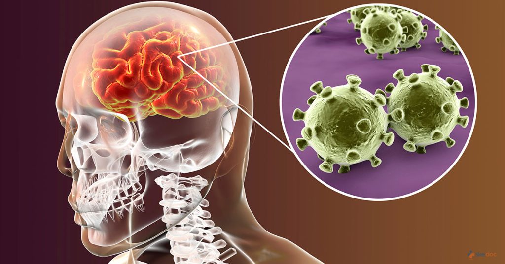
Type 1 diabetes Type 1 diabetes, once known as juvenile diabetes or insulin-dependent diabetes, is a chronic condition in which the pancreas produces little or no insulin. Insulin is a hormone needed to allow sugar (glucose) to enter cells to produce energy. Different factors, including genetics and some viruses, may contribute to type 1 diabetes. Although type 1 diabetes usually appears during childhood or adolescence, it can develop in adults. Despite active research, type 1 diabetes has no cure. Treatment focuses on managing blood sugar levels with insulin, diet and lifestyle to prevent complications. Causes of Type 1 diabetes The exact cause of type 1 diabetes is unknown. Usually, the body’s own immune system  which normally fights harmful bacteria and viruses  mistakenly destroys the insulin-producing (islet, or islets of Langerhans) cells in the pancreas. Other possible causes include: The role of insulin Once a significant number of islet cells are destroyed, you’ll produce little or no insulin. Insulin is a hormone that comes from a gland situated behind and below the stomach (pancreas). The role of glucose Glucose a sugar is a main source of energy for the cells that make up muscles and other tissues. When your glucose levels are low, such as when you haven’t eaten in a while, the liver breaks down the stored glycogen into glucose to keep your glucose levels within a normal range. In type 1 diabetes, there’s no insulin to let glucose into the cells, so sugar builds up in your bloodstream. This can cause life-threatening complications. Symptoms of Type 1 diabetes Type 1 diabetes signs and symptoms can appear relatively suddenly and may include: When to see a doctor Consult your doctor if you notice any of the above signs and symptoms in you or your child. Risk factors Some known risk factors for type 1 diabetes include: Family history.  Anyone with a parent or sibling with type 1 diabetes has a slightly increased risk of developing the condition. Genetics.  The presence of certain genes indicates an increased risk of developing type 1 diabetes. Geography.  The incidence of type 1 diabetes tends to increase as you travel away from the equator. Age.  Although type 1 diabetes can appear at any age, it appears at two noticeable peaks. The first peak occurs in children between 4 and 7 years old, and the second is in children between 10 and 14 years old. Complications Over time, type 1 diabetes complications can affect major organs in your body, including heart, blood vessels, nerves, eyes and kidneys. Maintaining a normal blood sugar level can dramatically reduce the risk of many complications. Eventually, diabetes complications may be disabling or even life-threatening. Heart and blood vessel disease.  Diabetes dramatically increases your risk of various cardiovascular problems, including coronary artery disease with chest pain (angina), heart attack, stroke, narrowing of the arteries (atherosclerosis) and high blood pressure. Nerve damage (neuropathy).  Excess sugar can injure the walls of the tiny blood vessels (capillaries) that nourish your nerves, especially in the legs. This can cause tingling, numbness, burning or pain that usually begins at the tips of the toes or fingers and gradually spreads upward. Poorly controlled blood sugar could cause you to eventually lose all sense of feeling in the affected limbs. Damage to the nerves that affect the gastrointestinal tract can cause problems with nausea, vomiting, diarrhea or constipation. For men, erectile dysfunction may be an issue. Kidney damage (nephropathy).  The kidneys contain millions of tiny blood vessel clusters that filter waste from your blood. Diabetes can damage this delicate filtering system. Severe damage can lead to kidney failure or irreversible end-stage kidney disease, which requires dialysis or a kidney transplant. Eye damage.  Diabetes can damage the blood vessels of the retina (diabetic retinopathy), potentially causing blindness. Diabetes also increases the risk of other serious vision conditions, such as cataracts and glaucoma. Foot damage.  Nerve damage in the feet or poor blood flow to the feet increases the risk of various foot complications. Left untreated, cuts and blisters can become serious infections that may ultimately require toe, foot or leg amputation. Skin and mouth conditions.  Diabetes may leave you more susceptible to infections of the skin and mouth, including bacterial and fungal infections. Gum disease and dry mouth also are more likely. Pregnancy complications.  High blood sugar levels can be dangerous for both the mother and the baby. The risk of miscarriage, stillbirth and birth defects increases when diabetes isn’t well-controlled. For the mother, diabetes increases the risk of diabetic ketoacidosis, diabetic eye problems (retinopathy), pregnancy-induced high blood pressure and preeclampsia. Prevention There’s no known way to prevent type 1 diabetes. But researchers are working on preventing the disease or further destruction of the islet cells in people who are newly diagnosed. Ask your doctor if you might be eligible for one of these clinical trials, but carefully weigh the risks and benefits of any treatment available in a trial. Diagnosis Diagnostic tests include: Glycated hemoglobin (A1C) test.  This blood test indicates your average blood sugar level for the past two to three months. It measures the percentage of blood sugar attached to the oxygen-carrying protein in red blood cells (hemoglobin). The higher your blood sugar levels, the more hemoglobin you’ll have with sugar attached. An A1C level of 6.5 percent or higher on two separate tests indicates diabetes. If the A1C test isn’t available, or if you have certain conditions that can make the A1C test inaccurate such as pregnancy or an uncommon form of hemoglobin (hemoglobin variant) your doctor may use these tests: Random blood sugar test.  A blood sample will be taken at a random time and may be confirmed by repeat testing. Blood sugar values are expressed in milligrams per deciliter (mg/dL) or millimoles per liter (mmol/L). Regardless of when you last ate, a random blood sugar level of 200 mg/dL (11.1 mmol/L) or higher suggests diabetes, especially when coupled with any of the signs and symptoms of diabetes, such as frequent urination and

Type 1 diabetes Read More »

Type 2 diabetes in children

Type 2 diabetes in children Type 2 diabetes in children is a chronic disease that affects the way your child’s body processes sugar (glucose). It’s important to manage your child’s diabetes because its long-term consequences can be disabling or even life-threatening. Type 2 diabetes is more commonly associated with adults. In fact, it used to be called adult-onset diabetes. But type 2 diabetes in children is on the rise, fueled largely by the obesity epidemic. There’s plenty you can do to help manage or prevent type 2 diabetes in children. Encourage your child to eat healthy foods, get plenty of physical activity and maintain a healthy weight. If diet and exercise aren’t enough to control type 2 diabetes in children, oral medication or insulin treatment may be needed. Causes of Type 2 diabetes in children The exact cause of type 2 diabetes is unknown. But family history and genetics appear to play an important role. Inactivity and excess fat especially abdominal fat also seem to be important factors. What is clear is that people with type 2 diabetes don’t process glucose properly anymore. As a result, sugar accumulates in the bloodstream instead of doing its normal job of fueling the cells that make up muscles and other tissues. Most of the glucose in people’s bodies comes from the food they eat. When food is digested, sugar enters the bloodstream. Moving sugar from the bloodstream to the body’s cells requires a hormone (insulin). Insulin comes from the pancreas, a gland located behind the stomach. The pancreas secretes insulin into the bloodstream after a person eats. As insulin circulates, it allows sugar to enter the cells and lowers the amount of sugar in the bloodstream. As the blood sugar level drops, so does the secretion of insulin from the pancreas. Type 2 diabetes develops when the body becomes resistant to insulin or when the pancreas stops making enough insulin. The resulting buildup of sugar in the bloodstream can cause life-threatening complications. Symptoms of Type 2 diabetes in children Type 2 diabetes in children may develop gradually. About 40 percent of children who have type 2 diabetes have no signs or symptoms and are diagnosed during routine physical exams. Other children might experience: Increased thirst and frequent urination.   Excess sugar building up in your child’s bloodstream pulls fluid from tissues. As a result your child might be thirsty and drink and urinate more than usual. Weight loss. Without the energy that sugar supplies, muscle tissues and fat stores simply shrink. However, weight loss is less common in children with type 2 diabetes than in children with type 1 diabetes. Fatigue.  Lack of sugar in your child’s cells might make him or her tired and lethargic. Blurred vision.  If your child’s blood sugar is too high, fluid may be pulled from the lenses of your child’s eyes. Your child might be unable to focus clearly. Slow-healing sores or frequent infections. Type 2 diabetes affects your child’s ability to heal and resist infections. When to see a doctor See your child’s doctor if you notice any of the signs or symptoms of type 2 diabetes. Undiagnosed, the disease can cause serious damage. Diabetes screening is recommended for all children and adolescents who are overweight and have at least two other risk factors for type 2 diabetes. Risk factors Researchers don’t fully understand why some children develop type 2 diabetes and others don’t, even if they have similar risk factors. However, it’s clear that certain factors increase the risk, including: Weight.  Being overweight is a primary risk factor for type 2 diabetes. The more fatty tissue children have  especially inside and between the muscle and skin around the abdomen  the more resistant their bodies cells become to insulin. The association between obesity and type 2 diabetes is even stronger in youth than in adults. Inactivity.  The less active your child is, the greater his or her risk of type 2 diabetes. Physical activity helps your child control his or her weight, uses glucose as energy, and makes your child’s cells more responsive to insulin. Family history.  Children’s risk of type 2 diabetes increases if they have a parent or sibling with the disease. Race. Although it’s unclear why, people of certain races mostly Africans develop type 2 diabetes. Age and sex.  Many children develop type 2 diabetes at the start of puberty. Adolescent girls are likelier to develop type 2 diabetes than are adolescent boys. Birth weight and gestational diabetes.  Low birth weight and being born to a mother who had gestational diabetes during the pregnancy are both associated with a higher risk of developing type 2 diabetes. Complications Type 2 diabetes can affect nearly every major organ in your child’s body, including the blood vessels, nerves, eyes and kidneys. The long-term complications of type 2 diabetes develop gradually. But eventually, diabetes complications may be disabling or even life-threatening. Complications of type 2 diabetes include: N.B – Keeping your child’s blood sugar level close to normal most of the time can dramatically reduce the risk of these complications. Prevention Healthy lifestyle choices can help prevent type 2 diabetes in children and its complications. And if your child already has type 2 diabetes, lifestyle changes can reduce the need for medications. Encourage your child to: Eat healthy foods.  Offer your child foods low in fat and calories. Focus on fruits, vegetables and whole grains. Strive for variety to prevent boredom. Get more physical activity.  Encourage your child to become active. Sign up for a sports team or dance lessons, or look for active things to do together. Better yet, make it a family affair.  The same lifestyle choices that can help prevent type 2 diabetes in children can do the same for adults. The best diet for a child with diabetes is also the best diet for the whole family. Diagnosis If diabetes is suspected, your child’s doctor will likely recommend a screening test. A diagnosis of type 2 diabetes in children generally requires abnormal

Type 2 diabetes in children Read More »

Vitiligo

Vitiligo Vitiligo is a disease that causes the loss of skin color in blotches. The extent and rate of color loss from vitiligo is unpredictable. It can affect the skin on any part of your body. It may also affect hair and the inside of the mouth. Normally, the color of hair and skin is determined by melanin. Vitiligo occurs when the cells that produce melanin die or stop functioning. Vitiligo affects people of all skin types, but it may be more noticeable in people with darker skin. The condition is not life-threatening or contagious. It can be stressful or make you feel bad about yourself. Treatment for vitiligo may restore color to the affected skin. But it does not prevent continued loss of skin color or a recurrence. Causes of Vitiligo Vitiligo occurs when pigment-producing cells (melanocytes) die or stop producing melanin the pigment that gives your skin, hair and eyes color. The involved patches of skin become lighter or white. Doctors don’t know why the cells fail or die. It may be related to: Symptoms of Vitiligo The main sign of vitiligo is patchy loss of skin color. Usually, the discoloration first shows on sun-exposed areas, such as the hands, feet, arms, face and lips. Vitiligo signs include: Depending on the type of vitiligo you have, the discolored patches may cover: Many parts of your body.  With this most common type, called generalized vitiligo, the discolored patches often progress similarly on corresponding body parts (symmetrically). Only one side or part of your body.  This type, called segmental vitiligo, tends to occur at a younger age, progress for a year or two, then stop. One or only a few areas of your body. This type is called localized (focal) vitiligo. It’s difficult to predict how your disease will progress.  Sometimes the patches stop forming without treatment. In most cases, pigment loss spreads and eventually involves most of your skin. Rarely, the skin gets its color back. When to see a doctor See your doctor if areas of your skin, hair or eyes lose coloring. Vitiligo has no cure. But treatment may help to stop or slow the discoloring process and return some color to your skin. Complications People with vitiligo may be at increased risk of: Diagnosis Medical history and exam If your doctor suspects you have vitiligo, he or she will ask about your medical history, examine you and try to rule out other medical problems, such as dermatitis or psoriasis. He or she may use a special lamp to shine ultraviolet light onto the skin to determine whether you have vitiligo. Skin biopsy and blood draw In addition to gathering your personal and family medical history and examining your skin, your doctor may: Treatment Many treatments are available to help restore skin color or even out skin tone. Results vary and are unpredictable. Some treatments have serious side effects. So your doctor may suggest that you first try improving the appearance of your skin by applying self-tanning products or makeup. If you and your doctor decide to treat your condition with a drug, surgery or therapy, the process may take many months to judge its effectiveness. And you may have to try more than one approach or a combination of approaches before you find the treatment that works best for you. Even if treatment is successful for a while, the results may not last or new patches may appear. Medications No drug can stop the process of vitiligo the loss of pigment cells (melanocytes).  But some drugs, used alone or with light therapy, can help restore some skin tone. Creams that control inflammation.  Applying a corticosteroid cream to affected skin may help return color, particularly if you start using it early in the disease. You may not see a change in your skin’s color for several months. This type of cream is effective and easy to use. But it can cause side effects, such as skin thinning or the appearance of streaks or lines on your skin. Milder forms of the drug may be prescribed for children and for people who have large areas of discolored skin. Medications that affect the immune system.  Ointments containing tacrolimus or pimecrolimus (calcineurin inhibitors) may be effective for people with small areas of depigmentation, especially on the face and neck. This treatment may have fewer side effects than corticosteroids and can be used with ultraviolet B (UVB) light. However, the Food and Drug Administration has warned about a possible link between these drugs and lymphoma and skin cancer. Therapies Combining psoralen and light therapy.  This treatment combines a plant-derived substance called psoralen with light therapy (photochemotherapy) to return color to the light patches. After you take psoralen by mouth or apply it to the affected skin, you’re exposed to ultraviolet A (UVA), UVB light or excimer light. These approaches tend to have better results than just medication or just light. You may need to repeat treatments up to three times a week for six to 12 months. Removing the remaining color (depigmentation).  This therapy may be an option if your vitiligo is widespread and other treatments haven’t worked. A depigmenting agent is applied to unaffected areas of skin. This gradually lightens it so that it blends with the discolored areas. The therapy is done once or twice a day for nine months or longer. Side effects can include redness, swelling, itching and dry skin. Depigmentation is permanent, and you’ll always be extremely sensitive to sunlight. Surgery Surgery may be an option for you if light therapy doesn’t work. Surgery can also be used with those therapies. The goal of the following techniques is to even out your skin tone by restoring color. Skin grafting.  In this procedure, your doctor removes very small sections of your normal, pigmented skin and attaches them to areas that have lost pigment. This procedure is sometimes used if you have small patches of vitiligo. Possible risks include infection, scarring, a cobblestone appearance, spotty color and

Vitiligo Read More »

Varicose veins

Varicose veins Varicose veins are twisted, enlarged veins. Any superficial vein may become varicosed, but the veins most commonly affected are those in your legs. That’s because standing and walking upright increases the pressure in the veins of your lower body. For many people, varicose veins and spider veins a common, mild variation of varicose veins are simply a cosmetic concern. For other people, varicose veins can cause aching pain and discomfort. Sometimes varicose veins lead to more-serious problems. Treatment may involve self-care measures or procedures by your doctor to close or remove veins. Causes of Varicose veins Weak or damaged valves can lead to varicose veins. Arteries carry blood from your heart to the rest of your tissues, and veins return blood from the rest of your body to your heart, so the blood can be recirculated. To return blood to your heart, the veins in your legs must work against gravity. Muscle contractions in your lower legs act as pumps, and elastic vein walls help blood return to your heart. Tiny valves in your veins open as blood flows toward your heart then close to stop blood from flowing backward. If these valves are weak or damaged, blood can flow backward and pool in the vein, causing the veins to stretch or twist. Symptoms of Varicose veins Varicose veins may not cause any pain. Signs you may have varicose veins include: When painful signs and symptoms occur, they may include: Spider veins are similar to varicose veins, but they’re smaller. Spider veins are found closer to the skin’s surface and are often red or blue. Spider veins occur on the legs, but can also be found on the face. They vary in size and often look like a spider’s web. When to see a doctor Self-care such as exercise, elevating your legs or wearing compression stockings can help you ease the pain of varicose veins and may prevent them from getting worse. But if you’re concerned about how your veins look and feel and self-care measures haven’t stopped your condition from getting worse, see your doctor. Risk factors These factors increase your risk of developing varicose veins: Age.  The risk of varicose veins increases with age. Aging causes wear and tear on the valves in your veins that help regulate blood flow. Eventually, that wear causes the valves to allow some blood to flow back into your veins where it collects instead of flowing up to your heart. Sex.  Women are more likely to develop the condition. Hormonal changes during pregnancy, premenstruation or menopause may be a factor because female hormones tend to relax vein walls. Hormone treatments, such as birth control pills, may increase your risk of varicose veins. Pregnancy. During pregnancy, the volume of blood in your body increases. This change supports the growing fetus, but also can produce an unfortunate side effect enlarged veins in your legs. Hormonal changes during pregnancy may also play a role. Family history.  If other family members had varicose veins, there’s a greater chance you will too. Obesity. Being overweight puts added pressure on your veins. Standing or sitting for long periods of time.  Your blood doesn’t flow as well if you’re in the same position for long periods. Complications Complications of varicose veins, although rare, can include: Ulcers. Painful ulcers may form on the skin near varicose veins, particularly near the ankles. A discolored spot on the skin usually begins before an ulcer forms. See your doctor immediately if you suspect you’ve developed an ulcer. Blood clots.  Occasionally, veins deep within the legs become enlarged. In such cases, the affected leg may become painful and swell. Any persistent leg pain or swelling warrants medical attention because it may indicate a blood clot a condition known medically as thrombophlebitis. Bleeding.  Occasionally, veins very close to the skin may burst. This usually causes only minor bleeding. But any bleeding requires medical attention. Prevention There’s no way to completely prevent varicose veins. But improving your circulation and muscle tone may reduce your risk of developing varicose veins or getting additional ones. The same measures you can take to treat the discomfort from varicose veins at home can help prevent varicose veins, including: Diagnosis To diagnose varicose veins, your doctor will do a physical exam, including looking at your legs while you’re standing to check for swelling. Your doctor may also ask you to describe any pain and aching in your legs. You also may need an ultrasound test to see if the valves in your veins are functioning normally or if there’s any evidence of a blood clot. In this noninvasive test, a technician runs a small hand-held device (transducer), about the size of a bar of soap, against your skin over the area of your body being examined. The transducer transmits images of the veins in your legs to a monitor, so a technician and your doctor can see them. Treatment Fortunately, treatment usually doesn’t mean a hospital stay or a long, uncomfortable recovery. Thanks to less invasive procedures, varicose veins can generally be treated on an outpatient basis. Ask your doctor if insurance will cover any of the cost of your treatment. If done for purely cosmetic reasons, you’ll likely have to pay for the treatment of varicose veins yourself. Self-care Self-care such as exercising, losing weight, not wearing tight clothes, elevating your legs, and avoiding long periods of standing or sitting can ease pain and prevent varicose veins from getting worse. Compression stockings Wearing compression stockings all day is often the first approach to try before moving on to other treatments. They steadily squeeze your legs, helping veins and leg muscles move blood more efficiently. The amount of compression varies by type and brand. You can buy compression stockings at most pharmacies and medical supply stores. Prescription-strength stockings also are available, and are likely covered by insurance if your varicose veins are causing symptoms. Additional treatments for more-severe varicose veins If you don’t respond to self-care or compression

Varicose veins Read More »

Umbilical hernia

Umbilical hernia An umbilical hernia occurs when part of your intestine sticks out through the opening in your abdominal muscles through which your umbilical cord passed before you were born. Umbilical hernias are common and typically harmless. They are most common in infants, but they can affect adults as well. In an infant, an umbilical hernia may be especially evident when the infant cries, causing the bellybutton to protrude. This is a classic sign of an umbilical hernia. Children’s umbilical hernias often close on their own in the first two years of life, though some remain open into the fifth year or longer. Umbilical hernias that appear during adulthood are more likely to need surgical repair. Causes of Umbilical hernia During gestation, the umbilical cord passes through a small opening in the baby’s abdominal muscles. The opening normally closes just after birth. If the muscles don’t join together completely in the midline of the abdominal wall, an umbilical hernia may appear at birth or later in life. In adults, too much abdominal pressure contributes to umbilical hernias. Causes of increased pressure in the abdomen include: Symptoms of Umbilical hernia An umbilical hernia creates a soft swelling or bulge near the navel (umbilicus). In babies who have an umbilical hernia, the bulge may be visible only when they cry, cough or strain. Umbilical hernias in children are usually painless. Umbilical hernias that appear during adulthood may cause abdominal discomfort. When to see a doctor If you suspect that your baby has an umbilical hernia, talk with the baby’s pediatrician. Seek emergency care if your baby has an umbilical hernia and: N.B – Similar guidelines apply to adults. Talk with your doctor if you have a bulge near your navel. Seek emergency care if the bulge becomes painful or tender. Prompt diagnosis and treatment can help prevent complications. Risk factors Umbilical hernias are most common in infants especially premature babies and those with low birth weights. In the United States, African American infants appear to have a slightly increased risk of umbilical hernias. The condition affects boys and girls equally. For adults, being overweight or having multiple pregnancies may increase the risk of developing an umbilical hernia. This type of hernia tends to be more common in women. Complications For children, complications of an umbilical hernia are rare. Complications can occur when the protruding abdominal tissue becomes trapped (incarcerated) and can no longer be pushed back into the abdominal cavity. This reduces the blood supply to the section of trapped intestine and can lead to umbilical pain and tissue damage. If the trapped portion of intestine is completely cut off from the blood supply (strangulated hernia), tissue death (gangrene) may occur. Infection may spread throughout the abdominal cavity, causing a life-threatening situation. Adults with umbilical hernias are somewhat more likely to experience incarceration or obstruction of the intestines. Emergency surgery is typically required to treat these complications. Diagnosis An umbilical hernia is diagnosed during a physical exam. Sometimes imaging studies such as an abdominal ultrasound or a CT scan are used to screen for complications. Treatment Most umbilical hernias in babies close on their own by age 1 or 2. Your doctor may even be able to push the bulge back into the abdomen during a physical exam. Don’t try this on your own, however. Although some people claim a hernia can be fixed by taping a coin down over the bulge, this “fix” doesn’t help and germs may accumulate under the tape, causing infection. For children, surgery is typically reserved for umbilical hernias that: During surgery, a small incision is made at the base of the bellybutton. The herniated tissue is returned to the abdominal cavity, and the opening in the abdominal wall is stitched closed. In adults, surgeons often use mesh to help strengthen the abdominal wall. Call us on : +27 82 0941 375   Email        : info@healthalert.co.za   Website   : www.healthalert.co.za   

Umbilical hernia Read More »

Ulcerative colitis

Ulcerative colitis Ulcerative colitis is an inflammatory bowel disease (IBD) that causes long-lasting inflammation and ulcers (sores) in your digestive tract. Ulcerative colitis affects the innermost lining of your large intestine (colon) and rectum. Symptoms usually develop over time, rather than suddenly. Ulcerative colitis can be debilitating and can sometimes lead to life-threatening complications. While it has no known cure, treatment can greatly reduce signs and symptoms of the disease and even bring about long-term remission. Causes of Ulcerative colitis The exact cause of ulcerative colitis remains unknown. Previously, diet and stress were suspected, but now doctors know that these factors may aggravate but don’t cause ulcerative colitis. One possible cause is an immune system malfunction. When your immune system tries to fight off an invading virus or bacterium, an abnormal immune response causes the immune system to attack the cells in the digestive tract, too. Heredity also seems to play a role in that ulcerative colitis is more common in people who have family members with the disease. However, most people with ulcerative colitis don’t have this family history. Symptoms of Ulcerative colitis Ulcerative colitis symptoms can vary, depending on the severity of inflammation and where it occurs. Signs and symptoms may include: N.B – Most people with ulcerative colitis have mild to moderate symptoms. The course of ulcerative colitis may vary, with some people having long periods of remission. Types of Ulcerative colitis Doctors often classify ulcerative colitis according to its location. Types of ulcerative colitis include: Ulcerative proctitis.  Inflammation is confined to the area closest to the anus (rectum), and rectal bleeding may be the only sign of the disease. This form of ulcerative colitis tends to be the mildest. Proctosigmoiditis.  Inflammation involves the rectum and sigmoid colon (lower end of the colon). Signs and symptoms include bloody diarrhea, abdominal cramps and pain, and an inability to move the bowels in spite of the urge to do so (tenesmus). Left-sided colitis.  Inflammation extends from the rectum up through the sigmoid and descending colon. Signs and symptoms include bloody diarrhea, abdominal cramping and pain on the left side, and unintended weight loss. Pancolitis.  Pancolitis often affects the entire colon and causes bouts of bloody diarrhea that may be severe, abdominal cramps and pain, fatigue, and significant weight loss. Acute severe ulcerative colitis.  This rare form of colitis affects the entire colon and causes severe pain, profuse diarrhea, bleeding, fever and inability to eat. When to see a doctor See your doctor if you experience a persistent change in your bowel habits or if you have signs and symptoms such as: Although ulcerative colitis usually isn’t fatal, it’s a serious disease that, in some cases, may cause lifethreatening complications. Risk factors Ulcerative colitis affects about the same number of women and men. Risk factors may include: Age.  Ulcerative colitis usually begins before the age of 30. But, it can occur at any age, and some people may not develop the disease until after age 60. Race or ethnicity.  Although whites have the highest risk of the disease, it can occur in any race. If you’re of Ashkenazi Jewish descent, your risk is even higher. Family history.  You’re at higher risk if you have a close relative, such as a parent, sibling or child, with the disease. Complications Possible complications of ulcerative colitis include: Diagnosis Your doctor will likely diagnose ulcerative colitis after ruling out other possible causes for your signs and symptoms. To help confirm a diagnosis of ulcerative colitis, you may have one or more of the following tests and procedures: Blood tests.  Your doctor may suggest blood tests to check for anemia a condition in which there aren’t enough red blood cells to carry adequate oxygen to your tissues or to check for signs of infection. Stool sample. White blood cells in your stool can indicate ulcerative colitis. A stool sample can also help rule out other disorders, such as infections caused by bacteria, viruses and parasites. Colonoscopy.  This exam allows your doctor to view your entire colon using a thin, flexible, lighted tube with an attached camera. During the procedure, your doctor can also take small samples of tissue (biopsy) for laboratory analysis. Sometimes a tissue sample can help confirm a diagnosis. Flexible sigmoidoscopy.  Your doctor uses a slender, flexible, lighted tube to examine the rectum and sigmoid, the last portion of your colon. If your colon is severely inflamed, your doctor may perform this test instead of a full colonoscopy. X-ray.  If you have severe symptoms, your doctor may use a standard X-ray of your abdominal area to rule out serious complications, such as a perforated colon. CT scan. A CT scan of your abdomen or pelvis may be performed if your doctor suspects a complication from ulcerative colitis. A CT scan may also reveal how much of the colon is inflamed. Computerized tomography (CT) enterography and magnetic resonance (MR) enterography.  Your doctor may recommend one of these noninvasive tests if he or she wants to exclude any inflammation in the small intestine. These tests are more sensitive for finding inflammation in the bowel than are conventional imaging tests. MR enterography is a radiation-free alternative. Treatment Ulcerative colitis treatment usually involves either drug therapy or surgery. Several categories of drugs may be effective in treating ulcerative colitis. The type you take will depend on the severity of your condition and location. The drugs that work well for some people may not work for others, so it may take time to find a medication that helps you. 5-aminosalicylic acid (5-ASA) 5-aminosalicylic acid (5-ASA) is often the first step in the treatment of ulcerative colitis. Examples of this type of medication include sulfasalazine (Azulfidine), mesalamine (Asacol HD, Delzicol, others), balsalazide (Colazal) and olsalazine (Dipentum). Which one you take, and whether it is taken by mouth or as an enema or suppository, depends on the area of your colon that’s affected. Corticosteroids These drugs, which include prednisone and budesonide (Uceris), are generally reserved for moderate to severe

Ulcerative colitis Read More »

Type 2 diabetes

Type 2 diabetes Type 2 diabetes is a chronic condition that affects the way your body metabolizes sugar (glucose) an important source of fuel for your body. With type 2 diabetes, your body either resists the effects of insulin a hormone that regulates the movement of sugar into your cells or doesn’t produce enough insulin to maintain normal glucose levels. Type 2 diabetes used to be known as adult-onset diabetes, but today more children are being diagnosed with the disorder, probably due to the rise in childhood obesity. There’s no cure for type 2 diabetes, but losing weight, eating well and exercising can help manage the disease. If diet and exercise aren’t enough to manage your blood sugar well, you may also need diabetes medications or insulin therapy. Causes of Type 2 diabetes Type 2 diabetes develops when the body becomes resistant to insulin or when the pancreas is unable to produce enough insulin. Exactly why this happens is unknown, although genetics and environmental factors, such as being overweight and inactive, seem to be contributing factors. How insulin works Insulin is a hormone that comes from the gland situated behind and below the stomach (pancreas). The role of glucose Glucose a sugar is a main source of energy for the cells that make up muscles and other tissues. When your glucose levels are low, such as when you haven’t eaten in a while, the liver breaks down stored glycogen into glucose to keep your glucose level within a normal range. In type 2 diabetes, this process doesn’t work well. Instead of moving into your cells, sugar builds up in your bloodstream. As blood sugar levels increase, the insulin-producing beta cells in the pancreas release more insulin, but eventually these cells become impaired and can’t make enough insulin to meet the body’s demands. In the much less common type 1 diabetes, the immune system mistakenly destroys the beta cells, leaving the body with little to no insulin. Symptoms of Type 2 diabetes Signs and symptoms of type 2 diabetes often develop slowly. In fact, you can have type 2 diabetes for years and not know it. Look for: When to see a doctor See your doctor if you notice type 2 diabetes symptoms. Risk factors Factors that may increase your risk of type 2 diabetes include: Weight.  Being overweight is a main risk factor for type 2 diabetes. However, you don’t have to be overweight to develop type 2 diabetes. Fat distribution.  If you store fat mainly in the abdomen, you have a greater risk of type 2 diabetes than if you store fat elsewhere, such as in your hips and thighs. Your risk of type 2 diabetes rises if you’re a man with a waist circumference above 40 inches (101.6 centimeters) or a woman with a waist that’s greater than 35 inches (88.9 centimeters). Inactivity.  The less active you are, the greater your risk of type 2 diabetes. Physical activity helps you control your weight, uses up glucose as energy and makes your cells more sensitive to insulin. Family history. The risk of type 2 diabetes increases if your parent or sibling has type 2 diabetes. Race. Although it’s unclear why, people of certain races including black, Hispanic, American Indian and AsianAmerican people are more likely to develop type 2 diabetes than white people are. Age.  The risk of type 2 diabetes increases as you get older, especially after age 45. That’s probably because people tend to exercise less, lose muscle mass and gain weight as they age. But type 2 diabetes is also increasing dramatically among children, adolescents and younger adults. Prediabetes.  Prediabetes is a condition in which your blood sugar level is higher than normal, but not high enough to be classified as diabetes. Left untreated, prediabetes often progresses to type 2 diabetes. Gestational diabetes.  If you developed gestational diabetes when you were pregnant, your risk of developing type 2 diabetes increases. If you gave birth to a baby weighing more than 9 pounds (4 kilograms), you’re also at risk of type 2 diabetes. Polycystic ovarian syndrome.  For women, having polycystic ovarian syndrome a common condition characterized by irregular menstrual periods, excess hair growth and obesity increases the risk of diabetes. Areas of darkened skin, usually in the armpits and neck. This condition often indicates insulin resistance. Complications Type 2 diabetes can be easy to ignore, especially in the early stages when you’re feeling fine. But diabetes affects many major organs, including your heart, blood vessels, nerves, eyes and kidneys. Controlling your blood sugar levels can help prevent these complications. Although long-term complications of diabetes develop gradually, they can eventually be disabling or even life-threatening. Some of the potential complications of diabetes include: Heart and blood vessel disease.  Diabetes dramatically increases the risk of heart disease, stroke, high blood pressure and narrowing of blood vessels (atherosclerosis). Nerve damage (neuropathy).  Excess sugar can cause tingling, numbness, burning or pain that usually begins at the tips of the toes or fingers and gradually spreads upward. Eventually, you may lose all sense of feeling in the affected limbs. Damage to the nerves that control digestion can cause problems with nausea, vomiting, diarrhea or constipation. For men, erectile dysfunction may be an issue. Kidney damage.  Diabetes can sometimes lead to kidney failure or irreversible end-stage kidney disease, which may require dialysis or a kidney transplant. Eye damage.  Diabetes increases the risk of serious eye diseases, such as cataracts and glaucoma, and may damage the blood vessels of the retina, potentially leading to blindness. Slow healing.  Left untreated, cuts and blisters can become serious infections, which may heal poorly. Severe damage might require toe, foot or leg amputation. Hearing impairment.  Hearing problems are more common in people with diabetes. Skin conditions.  Diabetes may leave you more susceptible to skin problems, including bacterial and fungal infections. Sleep apnea.  Obstructive sleep apnea is common in people with type 2 diabetes. Obesity may be the main contributing factor to both conditions. Treating sleep apnea may

Type 2 diabetes Read More »

Type 1 diabetes in children

Type 1 diabetes in children Type 1 diabetes in children is a condition in which your child’s body no longer produces an important hormone (insulin). Your child needs insulin to survive, so you’ll have to replace the missing insulin. Type 1 diabetes in children used to be known as juvenile diabetes or insulin-dependent diabetes. The diagnosis of type 1 diabetes in children can be overwhelming at first. Suddenly you and your child depending on his or her age must learn how to give injections, count carbohydrates and monitor blood sugar. Type 1 diabetes in children requires consistent care. But advances in blood sugar monitoring and insulin delivery have improved the daily management of the condition. Causes of Type 1 diabetes in children The exact cause of type 1 diabetes is unknown. But in most people with type 1 diabetes, the body’s immune system which normally fights harmful bacteria and viruses mistakenly destroys insulinproducing (islet) cells in the pancreas. Genetics and environmental factors appear to play a role in this process. Insulin performs the critical job of moving sugar (glucose) from the bloodstream to the body’s cells. Sugar enters the bloodstream when food is digested. Once the islet cells of the pancreas are destroyed, your child produces little or no insulin. As a result, glucose builds up in your child’s bloodstream, where it can cause life-threatening complications. Symptoms of Type 1 diabetes in children The signs and symptoms of type 1 diabetes in children usually develop quickly, over a period of weeks. These signs and symptoms include: Increased thirst and frequent urination.  Excess sugar building up in your child’s bloodstream pulls fluid from tissues. As a result your child might be thirsty and drink and urinate more than usual. A young, toilet-trained child might suddenly experience bed-wetting. Extreme hunger.  Without enough insulin to move sugar into your child’s cells, your child’s muscles and organs lack energy. This triggers intense hunger. Weight loss.  Despite eating more than usual to relieve hunger, your child may lose weight sometimes rapidly. Without the energy sugar supplies, muscle tissues and fat stores simply shrink. Unexplained weight loss is often the first sign of type 1 diabetes to be noticed in children. Fatigue.  Lack of sugar in your child’s cells might make him or her tired and lethargic. Irritability or behavior changes.  In addition to mood problems, your child might suddenly have a decline in performance at school. Fruity-smelling breath.  Burning fat instead of sugar produces certain substances (ketones) that can cause a fruity breath odor. Blurred vision.  If your child’s blood sugar is too high, fluid may be pulled from the lenses of your child’s eyes. Your child might be unable to focus clearly. Yeast infection.  Girls with type 1 diabetes may have a genital yeast infections. Babies can develop diaper rashes caused by yeast. When to see a doctor See your child’s doctor if you notice any of the signs or symptoms of type 1 diabetes. Risk factors Risk factors for type 1 diabetes in children include: Family history.  Anyone with a parent or siblings with type 1 diabetes has a slightly increased risk of developing the condition. Genetic susceptibility.  The presence of certain genes indicates an increased risk of developing type 1 diabetes. Race.  In the United States, type 1 diabetes is more common among non-Hispanic white children than among other races also European countries. Environmental risk factors might include: Certain viruses.  Exposure to various viruses may trigger the autoimmune destruction of the islet cells. Diet.  No specific dietary factor or nutrient in infancy has been shown to play a role in the development of type 1 diabetes. However, early intake of cow’s milk has been linked to an increased risk of type 1 diabetes, while breast-feeding might lower the risk. The timing of the introduction of cereal into a baby’s diet also may affect a child’s risk of type 1 diabetes. Complications Complications of type 1 diabetes develop gradually. If blood sugar levels aren’t well-controlled over a prolonged period of time, diabetes complications can eventually be disabling or even life-threatening. Complications can include: Heart and blood vessel disease.  Diabetes dramatically increases your child’s risk of developing conditions such as coronary artery disease with chest pain (angina), heart attack, stroke, narrowing of the arteries (atherosclerosis) and high blood pressure later in life. Nerve damage.  Excess sugar can injure the walls of the tiny blood vessels that nourish your child’s nerves, especially in the legs. This can cause tingling, numbness, burning or pain. Nerve damage usually happens gradually over a long period of time. Kidney damage.  Diabetes can damage the numerous tiny blood vessel clusters that filter waste from your child’s blood. Severe damage can lead to kidney failure or irreversible end-stage kidney disease, requiring dialysis or a kidney transplant. Eye damage.  Diabetes can damage the blood vessels of the retina, which may lead to poor vision and even possibly causing blindness. Diabetes can also lead to cataracts and a greater risk of glaucoma. Skin conditions.  Diabetes may leave your child more prone to skin problems, including bacterial infections, fungal infections and itching. Osteoporosis.  Diabetes may lead to lower than normal bone mineral density, increasing your child’s risk of osteoporosis as an adult. Prevention There’s currently no known way to prevent type 1 diabetes. Children who have a high risk of developing type 1 diabetes can be tested for antibodies associated with the disorder. But the presence of these antibodies doesn’t make diabetes inevitable. And there’s currently no known way to prevent type 1 diabetes if the antibodies are found. Researchers are working on preventing type 1 diabetes in people who have a high risk of developing the disease. Other research focuses on preventing further destruction of the islet cells in people who are newly diagnosed. While there’s nothing you could have done to prevent your child’s type 1 diabetes, you can help your child prevent its complications by: Diagnosis There are several blood tests for type 1 diabetes in children: Random blood sugar

Type 1 diabetes in children Read More »

Meningitis

Meningitis Meningitis is an inflammation of the membranes (meninges) surrounding your brain and spinal cord. The swelling from meningitis typically triggers symptoms such as headache, fever and a stiff neck. Most cases of meningitis in the World are caused by a viral infection, but bacterial, parasitic and fungal infections are other causes. Some cases of meningitis improve without treatment in a few weeks. Others can be life-threatening and require emergency antibiotic treatment. Seek immediate medical care if you suspect that someone has meningitis. Early treatment of bacterial meningitis can prevent serious complications. Causes of Meningitis Viral infections are the most common cause of meningitis, followed by bacterial infections and, rarely, fungal infections. Because bacterial infections can be life-threatening, identifying the cause is essential. Bacterial meningitis Bacteria that enter the bloodstream and travel to the brain and spinal cord cause acute bacterial meningitis. But it can also occur when bacteria directly invade the meninges. This may be caused by an ear or sinus infection, a skull fracture, or, rarely, after some surgeries. Several strains of bacteria can cause acute bacterial meningitis, most commonly: Streptococcus pneumoniae (pneumococcus).  This bacterium is the most common cause of bacterial meningitis in infants, young children and adults in the World. It more commonly causes pneumonia or ear or sinus infections. A vaccine can help prevent this infection. Neisseria meningitidis (meningococcus).  This bacterium is another leading cause of bacterial meningitis. These bacteria commonly cause an upper respiratory infection but can cause meningococcal meningitis when they enter the bloodstream. This is a highly contagious infection that affects mainly teenagers and young adults. It may cause local epidemics in college dormitories, boarding schools and military bases. A vaccine can help prevent infection. Haemophilus influenzae (haemophilus).  Haemophilus influenzae type b (Hib) bacterium was once the leading cause of bacterial meningitis in children. But new Hib vaccines have greatly reduced the number of cases of this type of meningitis. Listeria monocytogenes (listeria).  These bacteria can be found in unpasteurized cheeses, hot dogs and lunchmeats. Pregnant women, newborns, older adults and people with weakened immune systems are most susceptible. Listeria can cross the placental barrier, and infections in late pregnancy may be fatal to the baby. Viral meningitis Viral meningitis is usually mild and often clears on its own. Most cases in Zimbabwe it is caused by a group of viruses known as enteroviruses, which are most common in late summer and early fall. Viruses such as herpes simplex virus, HIV, mumps, West Nile virus and others also can cause viral meningitis. Chronic meningitis Slow-growing organisms (such as fungi and Mycobacterium tuberculosis) that invade the membranes and fluid surrounding your brain cause chronic meningitis. Chronic meningitis develops over two weeks or more. The signs and symptoms of chronic meningitis headaches, fever, vomiting and mental cloudiness  are similar to those of acute meningitis. Fungal meningitis Fungal meningitis is relatively uncommon and causes chronic meningitis. It may mimic acute bacterial meningitis. Fungal meningitis isn’t contagious from person to person. Cryptococcal meningitis is a common fungal form of the disease that affects people with immune deficiencies, such as AIDS. It’s lifethreatening if not treated with an antifungal medication. Other meningitis causes Meningitis can also result from noninfectious causes, such as chemical reactions, drug allergies, some types of cancer and inflammatory diseases such as sarcoidosis. Symptoms of Meningitis Early meningitis symptoms may mimic the flu (influenza). Symptoms may develop over several hours or over a few days. Possible signs and symptoms in anyone older than the age of 2 include: Signs in newborns Newborns and infants may show these signs: N.B – Infants with meningitis may be difficult to comfort, and may even cry harder when held. When to see a doctor Seek immediate medical care if you or someone in your family has meningitis symptoms, such as: N.B – Bacterial meningitis is serious, and can be fatal within days without prompt antibiotic treatment. Delayed treatment increases the risk of permanent brain damage or death. It’s also important to talk to your doctor if a family member or someone you work with has meningitis. You may need to take medications to prevent getting the infection. Risk factors Risk factors for meningitis include: Skipping vaccinations.  Risk rises for anyone who hasn’t completed the recommended childhood or adult vaccination schedule. Age.  Most cases of viral meningitis occur in children younger than age 5. Bacterial meningitis is common in those under age 20. Living in a community setting.  College students living in dormitories, personnel on military bases, and children in boarding schools and child care facilities are at greater risk of meningococcal meningitis. This is probably because the bacterium is spread by the respiratory route, and spreads quickly through large groups. Pregnancy.  Pregnancy increases the risk of listeriosis an infection caused by listeria bacteria, which may also cause meningitis. Listeriosis increases the risk of miscarriage, stillbirth and premature delivery. Compromised immune system.  AIDS, alcoholism, diabetes, use of immunosuppressant drugs and other factors that affect your immune system also make you more susceptible to meningitis. Having your spleen removed also increases your risk, and anyone without a spleen should get vaccinated to minimize that risk. Complications Meningitis complications can be severe. The longer you or your child has the disease without treatment, the greater the risk of seizures and permanent neurological damage, including: N.B – With prompt treatment, even patients with severe meningitis can have good recovery. Prevention Common bacteria or viruses that can cause meningitis can spread through coughing, sneezing, kissing, or sharing eating utensils, a toothbrush or a cigarette. These steps can help prevent meningitis: Wash your hands.  Careful hand-washing helps prevent the spread of germs. Teach children to wash their hands often, especially before eating and after using the toilet, spending time in a crowded public place or petting animals. Show them how to vigorously and thoroughly wash and rinse their hands. Practice good hygiene. Don’t share drinks, foods, straws, eating utensils, lip balms or toothbrushes with anyone else. Teach children and teens to avoid sharing

Meningitis Read More »

Infant jaundice

Infant jaundice Infant jaundice is yellow discoloration of a newborn baby’s skin and eyes. Infant jaundice occurs because the baby’s blood contains an excess of bilirubin, a yellow pigment of red blood cells. Infant jaundice is a common condition, particularly in babies born before 38 weeks’ gestation (preterm babies) and some breast-fed babies. Infant jaundice usually occurs because a baby’s liver isn’t mature enough to get rid of bilirubin in the bloodstream. In some babies, an underlying disease may cause infant jaundice. Most infants born between 35 weeks’ gestation and full term need no treatment for jaundice. Rarely, an unusually high blood level of bilirubin can place a newborn at risk of brain damage, particularly in the presence of certain risk factors for severe jaundice. Causes of Infant jaundice Excess bilirubin (hyperbilirubinemia) is the main cause of jaundice. Bilirubin, which is responsible for the yellow color of jaundice, is a normal part of the pigment released from the breakdown of “used” red blood cells. Newborns produce more bilirubin than adults do because of greater production and faster breakdown of red blood cells in the first few days of life. Normally, the liver filters bilirubin from the bloodstream and releases it into the intestinal tract. A newborn’s immature liver often can’t remove bilirubin quickly enough, causing an excess of bilirubin. Jaundice due to these normal newborn conditions is called physiologic jaundice, and it typically appears on the second or third day of life. Other causes An underlying disorder may cause infant jaundice. In these cases, jaundice often appears much earlier or much later than does the more common form of infant jaundice. Diseases or conditions that can cause jaundice include: Risk factors Major risk factors for jaundice, particularly severe jaundice that can cause complications, include: Premature birth.  A baby born before 38 weeks of gestation may not be able to process bilirubin as quickly as full-term babies do. Premature babies also may feed less and have fewer bowel movements, resulting in less bilirubin eliminated through stool. Significant bruising during birth.  Newborns who become bruised during delivery gets bruises from the delivery may have higher levels of bilirubin from the breakdown of more red blood cells. Blood type.  If the mother’s blood type is different from her baby’s, the baby may have received antibodies through the placenta that cause abnormally rapid breakdown of red blood cells. Breast-feeding.  Breast-fed babies, particularly those who have difficulty nursing or getting enough nutrition from breast-feeding, are at higher risk of jaundice. Dehydration or a low caloric intake may contribute to the onset of jaundice. However, because of the benefits of breast-feeding, experts still recommend it. It’s important to make sure your baby gets enough to eat and is adequately hydrated. Symptoms of Infant jaundice Yellowing of the skin and the whites of the eyes the main sign of infant jaundice usually appears between the second and fourth day after birth. To check for infant jaundice, press gently on your baby’s forehead or nose. If the skin looks yellow where you pressed, it’s likely your baby has mild jaundice. If your baby doesn’t have jaundice, the skin color should simply look slightly lighter than its normal color for a moment. Examine your baby in good lighting conditions, preferably in natural daylight. When to see a doctor Most hospitals have a policy of examining babies for jaundice before discharge. Pediatrics recommends that newborns be examined for jaundice during routine medical checks and at least every eight to 12 hours while in the hospital. Your baby should be examined for jaundice between the third and seventh day after birth, when bilirubin levels usually peak. If your baby is discharged earlier than 72 hours after birth, make a followup appointment to look for jaundice within two days of discharge. The following signs or symptoms may indicate severe jaundice or complications from excess bilirubin. Call your doctor if: Complications High levels of bilirubin that cause severe jaundice can result in serious complications if not treated. Acute bilirubin encephalopathy Bilirubin is toxic to cells of the brain. If a baby has severe jaundice, there’s a risk of bilirubin passing into the brain, a condition called acute bilirubin encephalopathy. Prompt treatment may prevent significant lasting damage. Signs of acute bilirubin encephalopathy in a baby with jaundice include: N.B – Kernicterus is the syndrome that occurs if acute bilirubin encephalopathy causes permanent damage to the brain.  Kernicterus may result in: Prevention The best preventive of infant jaundice is adequate feeding. Breast-fed infants should have eight to 12 feedings a day for the first several days of life. Formula-fed infants usually should have 1 to 2 ounces (about 30 to 60 milliliters) of formula every two to three hours for the first week. Diagnosis Your doctor will likely diagnose infant jaundice on the basis of your baby’s appearance. However, it’s still necessary to measure the level of bilirubin in your baby’s blood. The level of bilirubin (severity of jaundice) will determine the course of treatment. Tests to detect jaundice and measure bilirubin include: Treatment Mild infant jaundice often disappears on its own within two or three weeks. For moderate or severe jaundice, your baby may need to stay longer in the newborn nursery or be readmitted to the hospital. Treatments to lower the level of bilirubin in your baby’s blood may include: Light therapy (phototherapy).  Your baby may be placed under a special lamp that emits light in the blue-green spectrum. The light changes the shape and structure of bilirubin molecules in such a way that they can be excreted in both the urine and stool. During treatment, your baby will wear only a diaper and protective eye patches. Light therapy may be supplemented with the use of a light-emitting pad or mattress. Intravenous immunoglobulin (IVIg).  Jaundice may be related to blood type differences between mother and baby. This condition results in the baby carrying antibodies from the mother that contribute to the rapid breakdown of the baby’s red blood cells. Intravenous transfusion of

Infant jaundice Read More »