Uncategorized

Trichomoniasis

 Trichomoniasis Trichomoniasis is a common sexually transmitted infection caused by a parasite. In women, trichomoniasis can cause a foul-smelling vaginal discharge, genital itching and painful urination. Men who have trichomoniasis typically have no symptoms. Pregnant women who have trichomoniasis might be at higher risk of delivering their babies prematurely. To prevent reinfection with the organism that causes trichomoniasis, both partners should be treated. The most common treatment for trichomoniasis involves taking one megadose of metronidazole (Flagyl) or tinidazole (Tindamax). You can reduce your risk of infection by using condoms correctly every time you have sex. Causes of Trichomoniasis Trichomoniasis is caused by a one-celled protozoan, a type of tiny parasite that travels between people during sexual intercourse. The incubation period between exposure and infection is unknown, but it’s thought to range from five to 28 days. Symptoms of  Trichomoniasis Many women and most men with trichomoniasis have no symptoms, at least not at first. Trichomoniasis signs and symptoms for women include: N.B -Trichomoniasis rarely causes symptoms in men.  When men do have signs and symptoms, however, they might include: When to see a doctor See your doctor if you have a foul-smelling vaginal discharge or if you have pain with urination or sexual intercourse. Risk factors Risk factors include having: Complications Pregnant women who have trichomoniasis might:  Prevention As with other sexually transmitted infections, the only way to prevent trichomoniasis is to abstain from sex. To lower your risk, use condoms correctly every time you have sex. Diagnosis The diagnosis of trichomoniasis can be confirmed by looking at a sample of vaginal fluid for women or urine for men under a microscope. Growing a culture used to be the way to diagnose trichomoniasis, but newer, faster tests, such as rapid antigen tests and nucleic acid amplification, are more common now. Treatment The most common treatment for trichomoniasis, even for pregnant women, is to swallow one megadose of either metronidazole (Flagyl) or tinidazole (Tindamax). In some cases, your doctor might recommend a lower dose of metronidazole two times a day for seven days. Both you and your partner need treatment. And you need to avoid sexual intercourse until the infection is cured, which takes about a week. Don’t drink alcohol for 24 hours after taking metronidazole or 72 hours after taking tinidazole, because it can cause severe nausea and vomiting. Your doctor will likely want to retest you for trichomoniasis from two weeks to three months after treatment to be sure you haven’t been reinfected. N.B – Untreated, trichomoniasis can last for months to years. Visit us on : www.healthalert.co.za    Calls us on : +27 82 0941 375   Email us on : info@healthalert.co.za  

Trichomoniasis Read More »

Cataracts

Cataracts A cataract is a clouding of the normally clear lens of your eye. For people who have cataracts, seeing through cloudy lenses is a bit like looking through a frosty or fogged-up window. Clouded vision caused by cataracts can make it more difficult to read, drive a car (especially at night) or see the expression on a friend’s face. Most cataracts develop slowly and don’t disturb your eyesight early on. But with time, cataracts will eventually interfere with your vision. At first, stronger lighting and eyeglasses can help you deal with cataracts. But if impaired vision interferes with your usual activities, you might need cataract surgery. Fortunately, cataract surgery is generally a safe, effective procedure. Causes of cataract Most cataracts develop when aging or injury changes the tissue that makes up your eye’s lens. Some inherited genetic disorders that cause other health problems can increase your risk of cataracts. Cataracts can also be caused by other eye conditions, past eye surgery or medical conditions such as diabetes. Long-term use of steroid medications, too, can cause cataracts to develop. Symptoms of cataract Signs and symptoms of cataracts include: N.B – At first, the cloudiness in your vision caused by a cataract may affect only a small part of the eye’s lens and you may be unaware of any vision loss. As the cataract grows larger, it clouds more of your lens and distorts the light passing through the lens. This may lead to more noticeable symptoms. When to see a doctor? Make an appointment for an eye exam if you notice any changes in your vision. If you develop sudden vision changes, such as double vision or flashes of light, sudden eye pain, or sudden headache, see your doctor right away. How a cataract forms The lens, where cataracts form, is positioned behind the colored part of your eye (iris). The lens focuses light that passes into your eye, producing clear, sharp images on the retina  the lightsensitive membrane in the eye that functions like the film in a camera. As you age, the lenses in your eyes become less flexible, less transparent and thicker. Age-related and other medical conditions cause tissues within the lens to break down and clump together, clouding small areas within the lens. As the cataract continues to develop, the clouding becomes denser and involves a bigger part of the lens. A cataract scatters and blocks the light as it passes through the lens, preventing a sharply defined image from reaching your retina. As a result, your vision becomes blurred. Cataracts generally develop in both eyes, but not evenly. The cataract in one eye may be more advanced than the other, causing a difference in vision between eyes. Types of cataracts Cataract types include: Cataracts affecting the center of the lens (nuclear cataracts).  A nuclear cataract may at first cause more nearsightedness or even a temporary improvement in your reading vision. But with time, the lens gradually turns more densely yellow and further clouds your vision. As the cataract slowly progresses, the lens may even turn brown.  Advanced yellowing or browning of the lens can lead to difficulty distinguishing between shades of color. Cataracts that affect the edges of the lens (cortical cataracts).  A cortical cataract begins as whitish, wedge-shaped opacities or streaks on the outer edge of the lens cortex. As it slowly progresses, the streaks extend to the center and interfere with light passing through the center of the lens. Cataracts that affect the back of the lens (posterior subcapsular cataracts).  A posterior subcapsular cataract starts as a small, opaque area that usually forms near the back of the lens, right in the path of light. A posterior subcapsular cataract often interferes with your reading vision, reduces your vision in bright light, and causes glare or halos around lights at night. These types of cataracts tend to progress faster than other types do. Cataracts you’re born with (congenital cataracts).  Some people are born with cataracts or develop them during childhood. These cataracts may be genetic, or associated with an intrauterine infection or trauma. These cataracts also may be due to certain conditions, such as myotonic dystrophy, galactosemia, neurofibromatosis type 2 or rubella. Congenital cataracts don’t always affect vision, but if they do they’re usually removed soon after detection. Risk factors Factors that increase your risk of cataracts include: Prevention No studies have proved how to prevent cataracts or slow the progression of cataracts. But doctors think several strategies may be helpful, including: Have regular eye examinations.  Eye examinations can help detect cataracts and other eye problems at their earliest stages. Ask your doctor how often you should have an eye examination. Quit smoking.  Ask your doctor for suggestions about how to stop smoking. Medications, counseling and other strategies are available to help you. Manage other health problems.  Follow your treatment plan if you have diabetes or other medical conditions that can increase your risk of cataracts. Choose a healthy diet that includes plenty of fruits and vegetables.  Adding a variety of colorful fruits and vegetables to your diet ensures that you’re getting many vitamins and nutrients. Fruits and vegetables have many antioxidants, which help maintain the health of your eyes. Studies haven’t proved that antioxidants in pill form can prevent cataracts.  But, a large population study recently showed that a healthy diet rich in vitamins and minerals was associated with a reduced risk of developing cataracts. Fruits and vegetables have many proven health benefits and are a safe way to increase the amount of minerals and vitamins in your diet. Wear sunglasses.  Ultraviolet light from the sun may contribute to the development of cataracts. Wear sunglasses that block ultraviolet B (UVB) rays when you’re outdoors. Reduce alcohol use.  Excessive alcohol use can increase the risk of cataracts. Diagnosis To determine whether you have a cataract, your doctor will review your medical history and symptoms, and perform an eye examination. Your doctor may conduct several tests, including: Visual acuity test.  A visual acuity test

Cataracts Read More »

Erectile dysfunction

Erectile dysfunction Erectile dysfunction (impotence) is the inability to get and keep an erection firm enough for sex. Having erection trouble from time to time isn’t necessarily a cause for concern. If erectile dysfunction is an ongoing issue, however, it can cause stress, affect your self-confidence and contribute to relationship problems. Problems getting or keeping an erection can also be a sign of an underlying health condition that needs treatment and a risk factor for heart disease. If you’re concerned about erectile dysfunction, talk to your doctor even if you’re embarrassed. Sometimes, treating an underlying condition is enough to reverse erectile dysfunction. In other cases, medications or other direct treatments might be needed. Causes of Erectile dysfunction Male sexual arousal is a complex process that involves the brain, hormones, emotions, nerves, muscles and blood vessels. Erectile dysfunction can result from a problem with any of these. Likewise, stress and mental health concerns can cause or worsen erectile dysfunction. Sometimes a combination of physical and psychological issues causes erectile dysfunction. For instance, a minor physical condition that slows your sexual response might cause anxiety about maintaining an erection. The resulting anxiety can lead to or worsen erectile dysfunction. Physical causes of erectile dysfunction In many cases, erectile dysfunction is caused by something physical. Common causes include: The brain plays a key role in triggering the series of physical events that cause an erection, starting with feelings of sexual excitement. A number of things can interfere with sexual feelings and cause or worsen erectile dysfunction. These include: Symptoms of Erectile dysfunction Erectile dysfunction symptoms might include persistent: When to see a doctor? A family doctor is a good place to start when you have erectile problems. See your doctor if: Risk factors As you get older, erections might take longer to develop and might not be as firm. You might need more direct touch to your penis to get and keep an erection. Various risk factors can contribute to erectile dysfunction, including: Complications Complications resulting from erectile dysfunction can include: Prevention The best way to prevent erectile dysfunction is to make healthy lifestyle choices and to manage any existing health conditions. For example: Diagnosis For many men, a physical exam and answering questions (medical history) are all that’s needed for a doctor to diagnose erectile dysfunction and recommend a treatment. If you have chronic health conditions or your doctor suspects that an underlying condition might be involved, you might need further tests or a consultation with a specialist. Tests for underlying conditions might include: Physical exam.  This might include careful examination of your penis and testicles and checking your nerves for sensation. Blood tests. A sample of your blood might be sent to a lab to check for signs of heart disease, diabetes, low testosterone levels and other health conditions. Urine tests (urinalysis). Like blood tests, urine tests are used to look for signs of diabetes and other underlying health conditions. Ultrasound. This test is usually performed by a specialist in an office. It involves using a wandlike device (transducer) held over the blood vessels that supply the penis. It creates a video image to let your doctor see if you have blood flow problems. This test is sometimes done in combination with an injection of medications into the penis to stimulate blood flow and produce an erection. Psychological exam. Your doctor might ask questions to screen for depression and other possible psychological causes of erectile dysfunction. Treatment The first thing your doctor will do is to make sure you’re getting the right treatment for any health conditions that could be causing or worsening your erectile dysfunction. Depending on the cause and severity of your erectile dysfunction and any underlying health conditions, you might have various treatment options. Your doctor can explain the risks and benefits of each treatment and will consider your preferences. Your partner’s preferences also might play a role in your treatment choices. Oral medications Oral medications are a successful erectile dysfunction treatment for many men. They include: N.B – All four medications enhance the effects of nitric oxide a natural chemical your body produces that relaxes muscles in the penis. This increases blood flow and allows you to get an erection in response to sexual stimulation. Taking one of these tablets will not automatically produce an erection. Sexual stimulation is needed first to cause the release of nitric oxide from your penile nerves. These medications amplify that signal, allowing some men to function normally. Oral erectile dysfunction medications are not aphrodisiacs, will not cause excitement and are not needed in men who get normal erections. The medications vary in dosage, how long they work and side effects. Possible side effects include flushing, nasal congestion, headache, visual changes, backache and stomach upset. Your doctor will consider your particular situation to determine which medication might work best. These medications might not treat your erectile dysfunction immediately. You might need to work with your doctor to find the right medication and dosage for you. Before taking any medication for erectile dysfunction, including over-the-counter supplements and herbal remedies, get your doctor’s OK. Medications for erectile dysfunction do not work in all men and might be less effective in certain conditions, such as after prostate surgery or if you have diabetes. Some medications might also be dangerous if you: Other medications Other medications for erectile dysfunction include: Alprostadil self-injection. With this method, you use a fine needle to inject alprostadil (Caverject Impulse, Edex) into the base or side of your penis. In some cases, medications generally used for other conditions are used for penile injections on their own or in combination. Examples include papaverine, alprostadil and phentolamine. Often these combination medications are known as bimix (if two medications are included) or trimix (if three are included). Each injection is dosed to create an erection lasting no longer than an hour. Because the needle used is very fine, pain from the injection site is usually minor. Side effects can include mild bleeding from

Erectile dysfunction Read More »

Constipation in children

Constipation in children Constipation in children is a common problem. A constipated child has infrequent bowel movements or hard, dry stools. Common causes include early toilet training and changes in diet. Fortunately, most cases of constipation in children are temporary. Encouraging your child to make simple dietary changes such as eating more fiber-rich fruits and vegetables and drinking more water can go a long way toward alleviating constipation. If your child’s doctor approves, it may be possible to treat a child’s constipation with laxatives. Symptoms Constipation in children Signs and symptoms of constipation in children may include: If your child fears that having a bowel movement will hurt, he or she may try to avoid it. You may notice your child crossing his or her legs, clenching his or her buttocks, twisting his or her body, or making faces when attempting to hold stool. When to see a doctor Constipation in children usually isn’t serious. However, chronic constipation may lead to complications or signal an underlying condition. Take your child to a doctor if the constipation lasts longer than two weeks or is accompanied by: Causes Constipation in children Constipation most commonly occurs when waste or stool moves too slowly through the digestive tract, causing the stool to become hard and dry. Many factors can contribute to constipation in children, including : Withholding.  Your child may ignore the urge to have a bowel movement because he or she is afraid of the toilet or doesn’t want to take a break from play. Some children withhold when they’re away from home because they’re uncomfortable using public toilets. Painful bowel movements caused by large, hard stools also may lead to withholding.  If it hurts to poop, your child may try to avoid a repeat of the distressing experience. Toilet training issues.  If you begin toilet training too soon, your child may rebel and hold in stool. If toilet training becomes a battle of wills, a voluntary decision to ignore the urge to poop can quickly become an involuntary habit that’s tough to change. Changes in diet.  Not enough fiber-rich fruits and vegetables or fluid in your child’s diet may cause constipation. One of the more common times for children to become constipated is when they’re switching from an all-liquid diet to one that includes solid foods. Changes in routine.  Any changes in your child’s routine such as travel, hot weather or stress can affect bowel function. Children are also more likely to experience constipation when they first start school outside of the home. Medications.  Certain antidepressants and various other drugs can contribute to constipation. Cow’s milk allergy. An allergy to cow’s milk or consuming too many dairy products (cheese and cow’s milk) sometimes leads to constipation. Family history. Children who have family members who have experienced constipation are more likely to develop constipation. This may be due to shared genetic or environmental factors. Medical conditions.  Rarely, constipation in children indicates an anatomic malformation, a metabolic or digestive system problem, or another underlying condition. Risk factors Constipation in children Constipation in children is more likely to affect kids who: Complications Constipation in children  Although constipation in children can be uncomfortable, it usually isn’t serious. If constipation becomes chronic, however, complications may include: Prevention Constipation in children  To help prevent constipation in children: Offer your child high-fiber foods.   A diet rich in fiber can help your child’s body form soft, bulky stool. Serve your child more highfiber foods, such as fruits, vegetables, beans, and whole-grain cereals and breads. If your child isn’t used to a high-fiber diet, start by adding just several grams of fiber a day to prevent gas and bloating. The recommended intake for dietary fiber is 14 grams for every 1,000 calories in your child’s diet. For younger children, this translates to an intake of about 20 grams of dietary fiber a day. For adolescent girls and young women, it’s 29 grams a day. And for adolescent boys and young men, it’s 38 grams a day. Encourage your child to drink plenty of fluids. Water is often the best. Promote physical activity. Regular physical activity helps stimulate normal bowel function. Create a toilet routine. Regularly set aside time after meals for your child to use the toilet. If necessary, provide a footstool so that your child is comfortable sitting on the toilet and has enough leverage to release a stool. Remind your child to heed nature’s call.  Some children get so wrapped up in play that they ignore the urge to have a bowel movement. If such delays occur often, they can contribute to constipation. Be supportive.  Reward your child’s efforts, not results. Give children small rewards for trying to move their bowel. Review medications.  If your child is taking a medication that causes constipation, ask his or her doctor about other options. Diagnosis constipation in children Your child’s doctor will: Gather a complete medical history.  Your child’s doctor will ask you about your child’s past illnesses. He or she will also likely ask you about your child’s diet and physical activity patterns. Conduct a physical exam.  Your child’s physical exam will likely include placing a gloved finger into your child’s anus to check for abnormalities or the presence of impacted stool. Stool found in the rectum may be tested for blood. More-extensive testing is usually reserved for only the most severe cases of constipation. If necessary, these tests may include: Abdominal X-ray.  This standard X-ray test allows your child’s doctor to see if there are any blockages in your child’s abdomen. Anorectal manometry or motility test. In this test, a thin tube called a catheter is placed in the rectum to measure the coordination of the muscles your child uses to pass stool. Barium enema X-ray.  In this test, the lining of the bowel is coated with a contrast dye (barium) so that the rectum, colon and sometimes part of the small intestine can be clearly seen on an X-ray. Rectal biopsy.  In this test, a small sample

Constipation in children Read More »

Ganglion cyst

Ganglion cyst Ganglion cysts are noncancerous lumps that most commonly develop along the tendons or joints of your wrists or hands. They also may occur in the ankles and feet. Ganglion cysts are typically round or oval and are filled with a jellylike fluid. Small ganglion cysts can be pea-sized, while larger ones can be around an inch (2.5 centimeters) in diameter. Ganglion cysts can be painful if they press on a nearby nerve. Their location can sometimes interfere with joint movement. If your ganglion cyst is causing you problems, your doctor may suggest trying to drain the cyst with a needle. Removing the cyst surgically also is an option. But if you have no symptoms, no treatment is necessary. In many cases, the cysts go away on their own. Causes OF Ganglion cysts No one knows exactly what causes a ganglion cyst to develop. It grows out of a joint or the lining of a tendon, looking like a tiny water balloon on a stalk, and seems to occur when the tissue that surrounds a joint or a tendon bulges out of place. Inside the cyst is a thick lubricating fluid similar to that found in joints or around tendons Symptoms Ganglion cyst The lumps associated with ganglion cysts can be characterized by: The next most common locations are the ankles and feet. These cysts can occur near other joints as well.  Ganglion cysts are round or oval and usually measure less than an inch (2.5 centimeters) in diameter. Some are so small that they can’t be felt. The size of a cyst can fluctuate, often getting larger when you use that joint for repetitive motions. But if a cyst presses on a nerve — even if the cyst is too small to form a noticeable lump — it can cause pain, tingling, numbness or muscle weakness. When to see a doctor See your doctor if you experience a noticeable lump or pain in your wrist, hand, ankle or foot. He or she can make a diagnosis and determine whether you need treatment. Risk factors Factors that may increase your risk of ganglion cysts include: Your sex and age.  Ganglion cysts can develop in anyone, but they most commonly occur in women between the ages of 20 and 40. Osteoarthritis.  People who have wear-and-tear arthritis in the finger joints closest to their fingernails are at higher risk of developing ganglion cysts near those joints. Joint or tendon injury.  Joints or tendons that have been injured in the past are more likely to develop ganglion cysts. Diagnosis OF Ganglion cysts During the physical exam, your doctor may apply pressure to the cyst to test for tenderness or discomfort. He or she may try to shine a light through the cyst to determine if it’s a solid mass or filled with fluid. Your doctor might also recommend imaging tests such as X-rays, ultrasound or magnetic resonance imaging (MRI) to rule out other conditions, such as arthritis or a tumor. MRIs and ultrasounds also can locate hidden (occult) cysts. A ganglion cyst diagnosis may be confirmed by aspiration, a process in which your doctor uses a needle and syringe to draw out (aspirate) the fluid in the cyst. Fluid from a ganglion cyst will be thick and clear or translucent. Treatment Ganglion cysts Ganglion cysts are often painless, requiring no treatment. Your doctor may suggest a watch-and-wait approach. If the cyst is causing pain or interfering with joint movement, your doctor may recommend: Immobilization.  Because activity can cause the ganglion cyst to get larger, it may help to temporarily immobilize the area with a brace or splint. As the cyst shrinks, it may release the pressure on your nerves, relieving pain. Avoid long-term use of a brace or splint, which can cause the nearby muscles to weaken. Aspiration.  In this procedure, your doctor uses a needle to drain the fluid from the cyst. The cyst may recur. Surgery.  This may be an option if other approaches haven’t worked. During this procedure, the doctor removes the cyst and the stalk that attaches it to the joint or tendon. Rarely, the surgery can injure the surrounding nerves, blood vessels or tendons. And the cyst can recur, even after surgery. Lifestyle and home remedies To relieve pain, consider an over-the-counter pain reliever, such as ibuprofen (Advil, Motrin IB, others) or naproxen sodium (Aleve). In some cases, modifying your shoes or how you lace them can relieve the pain associated with ganglion cysts on your ankles or feet. Things not to do An old home remedy for a ganglion cyst consisted of “thumping” the cyst with a heavy object. This isn’t a good solution because the force of the blow can damage surrounding structures in your hand or foot. Also don’t try to “pop” the cyst yourself by puncturing it with a needle. This is unlikely to be effective and can lead to infection. Visit us on : www.healthalert.co.za    Calls us on : +27 82 0941 375   Email us on : info@healthalert.co.za  

Ganglion cyst Read More »

Gallstones

Gallstones Gallstones are hardened deposits of digestive fluid that can form in your gallbladder. Your gallbladder is a small, pear-shaped organ on the right side of your abdomen, just beneath your liver. The gallbladder holds a digestive fluid called bile that’s released into your small intestine. Gallstones range in size from as small as a grain of sand to as large as a golf ball. Some people develop just one gallstone, while others develop many gallstones at the same time. People who experience symptoms from their gallstones usually require gallbladder removal surgery. Gallstones that don’t cause any signs and symptoms typically don’t need treatment. Causes Gallstones It’s not clear what causes gallstones to form. Doctors think gallstones may result when: Your bile contains too much cholesterol.  Normally, your bile contains enough chemicals to dissolve the cholesterol excreted by your liver. But if your liver excretes more cholesterol than your bile can dissolve, the excess cholesterol may form into crystals and eventually into stones. Your bile contains too much bilirubin.  Bilirubin is a chemical that’s produced when your body breaks down red blood cells. Certain conditions cause your liver to make too much bilirubin, including liver cirrhosis, biliary tract infections and certain blood disorders. The excess bilirubin contributes to gallstone formation. Your gallbladder doesn’t empty correctly.  If your gallbladder doesn’t empty completely or often enough, bile may become very concentrated, contributing to the formation of gallstones. Types of gallstones Types of gallstones that can form in the gallbladder include: Cholesterol gallstones.  The most common type of gallstone, called a cholesterol gallstone, often appears yellow in color. These gallstones are composed mainly of undissolved cholesterol, but may contain other components. Pigment gallstones.  These dark brown or black stones form when your bile contains too much bilirubin. Symptoms Gallstones Gallstones may cause no signs or symptoms. If a gallstone lodges in a duct and causes a blockage, the resulting signs and symptoms may include: N.B – Gallstone pain may last several minutes to a few hours. When to see a doctor? Make an appointment with your doctor if you have any signs or symptoms that worry you. Seek immediate care if you develop signs and symptoms of a serious gallstone complication, such as: Factors that may increase your risk of gallstones include: Complications of Gallstones Complications of gallstones may include: Inflammation of the gallbladder.  A gallstone that becomes lodged in the neck of the gallbladder can cause inflammation of the gallbladder (cholecystitis). Cholecystitis can cause severe pain and fever. Blockage of the common bile duct.   Gallstones can block the tubes (ducts) through which bile flows from your gallbladder or liver to your small intestine. Severe pain, jaundice and bile duct infection can result. Blockage of the pancreatic duct.  The pancreatic duct is a tube that runs from the pancreas and connects to the common bile duct just before entering the duodenum. Pancreatic juices, which aid in digestion, flow through the pancreatic duct. N.B – A gallstone can cause a blockage in the pancreatic duct, which can lead to inflammation of the pancreas (pancreatitis). Pancreatitis causes intense, constant abdominal pain and usually requires hospitalization. Gallbladder cancer.  People with a history of gallstones have an increased risk of gallbladder cancer. But gallbladder cancer is very rare, so even though the risk of cancer is elevated, the likelihood of gallbladder cancer is still very small. Prevention of Gallstones You can reduce your risk of gallstones if you: Don’t skip meals.  Try to stick to your usual mealtimes each day. Skipping meals or fasting can increase the risk of gallstones. Lose weight slowly.  If you need to lose weight, go slow. Rapid weight loss can increase the risk of gallstones. Aim to lose 1 or 2 pounds (about 0.5 to 1 kilogram) a week. Eat more high-fiber foods.  Include more fiber-rich foods in your diet, such as fruits, vegetables and whole grains. Maintain a healthy weight.  Obesity and being overweight increase the risk of gallstones. Work to achieve a healthy weight by reducing the number of calories you eat and increasing the amount of physical activity you get. Once you achieve a healthy weight, work to maintain that weight by continuing your healthy diet and continuing to exercise. Diagnosis of Gallstones Tests and procedures used to diagnose gallstones and complications of gallstones include: Abdominal ultrasound.  This test is the one most commonly used to look for signs of gallstones. Abdominal ultrasound involves moving a device (transducer) back and forth across your stomach area. The transducer sends signals to a computer, which creates images that show the structures in your abdomen. Endoscopic ultrasound (EUS).  This procedure can help identify smaller stones that may be missed on an abdominal ultrasound. During EUS your doctor passes a thin, flexible tube (endoscope) through your mouth and through your digestive tract. A small ultrasound device (transducer) in the tube produces sound waves that create a precise image of surrounding tissue. Other imaging tests.  Additional tests may include oral cholecystography, a hepatobiliary iminodiacetic acid (HIDA) scan, computerized tomography (CT), magnetic resonance cholangiopancreatography (MRCP) or endoscopic retrograde cholangiopancreatography (ERCP). Gallstones discovered using ERCP can be removed during the procedure. Blood tests.  Blood tests may reveal infection, jaundice, pancreatitis or other complications caused by gallstones. Treatment of Gallstones Most people with gallstones that don’t cause symptoms will never need treatment. Your doctor will determine if treatment for gallstones is indicated based on your symptoms and the results of diagnostic testing. Your doctor may recommend that you be alert for symptoms of gallstone complications, such as intensifying pain in your upper right abdomen. If gallstone signs and symptoms occur in the future, you can have treatment. Treatment options for gallstones include: Surgery to remove the gallbladder (cholecystectomy).  Your doctor may recommend surgery to remove your gallbladder, since gallstones frequently recur. Once your gallbladder is removed, bile flows directly from your liver into your small intestine, rather than being stored in your gallbladder. You don’t need your gallbladder to live, and gallbladder removal doesn’t affect

Gallstones Read More »

Epilepsy

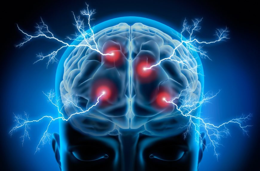
Epilepsy Epilepsy is a central nervous system (neurological) disorder in which brain activity becomes abnormal, causing seizures or periods of unusual behavior, sensations, and sometimes loss of awareness. Anyone can develop epilepsy. Epilepsy affects both males and females of all races, ethnic backgrounds and ages. Seizure symptoms can vary widely. Some people with epilepsy simply stare blankly for a few seconds during a seizure, while others repeatedly twitch their arms or legs. Having a single seizure doesn’t mean you have epilepsy. At least two unprovoked seizures are generally required for an epilepsy diagnosis. Treatment with medications or sometimes surgery can control seizures for the majority of people with epilepsy. Some people require lifelong treatment to control seizures, but for others, the seizures eventually go away. Some children with epilepsy may outgrow the condition with age. Symptoms of Epilepsy Because epilepsy is caused by abnormal activity in the brain, seizures can affect any process your brain coordinates. Seizure signs and symptoms may include: Symptoms vary depending on the type of seizure. In most cases, a person with epilepsy will tend to have the same type of seizure each time, so the symptoms will be similar from episode to episode. Doctors generally classify seizures as either focal or generalized, based on how the abnormal brain activity begins. Focal seizures When seizures appear to result from abnormal activity in just one area of your brain, they’re called focal (partial) seizures. These seizures fall into two categories: Focal seizures without loss of consciousness.  Once called simple partial seizures, these seizures don’t cause a loss of consciousness. They may alter emotions or change the way things look, smell, feel, taste or sound. They may also result in involuntary jerking of a body part, such as an arm or leg, and spontaneous sensory symptoms such as tingling, dizziness and flashing lights. Focal seizures with impaired awareness.  Once called complex partial seizures, these seizures involve a change or loss of consciousness or awareness. During a complex partial seizure, you may stare into space and not respond normally to your environment or perform repetitive movements, such as hand rubbing, chewing, swallowing or walking in circles. Symptoms of focal seizures may be confused with other neurological disorders, such as migraine, narcolepsy or mental illness. A thorough examination and testing are needed to distinguish epilepsy from other disorders. Generalized seizures Seizures that appear to involve all areas of the brain are called generalized seizures. Six types of generalized seizures exist. Absence seizures.  Absence seizures, previously known as petit mal seizures, often occur in children and are characterized by staring into space or subtle body movements such as eye blinking or lip smacking. These seizures may occur in clusters and cause a brief loss of awareness. Tonic seizures.  Tonic seizures cause stiffening of your muscles. These seizures usually affect muscles in your back, arms and legs and may cause you to fall to the ground. Atonic seizures.  Atonic seizures, also known as drop seizures, cause a loss of muscle control, which may cause you to suddenly collapse or fall down. Clonic seizures.  Clonic seizures are associated with repeated or rhythmic, jerking muscle movements. These seizures usually affect the neck, face and arms. Myoclonic seizures.  Myoclonic seizures usually appear as sudden brief jerks or twitches of your arms and legs. Tonic-clonic seizures.  Tonic-clonic seizures, previously known as grand mal seizures, are the most dramatic type of epileptic seizure and can cause an abrupt loss of consciousness, body stiffening and shaking, and sometimes loss of bladder control or biting your tongue. When to see a doctor? Seek immediate medical help if any of the following occurs: N.B – If you experience a seizure for the first time, seek medical advice. Causes of Epilepsy Epilepsy has no identifiable cause in about half the people with the condition. In the other half, the condition may be traced to various factors, including: Genetic influence.  Some types of epilepsy, which are categorized by the type of seizure you experience or the part of the brain that is affected, run in families. In these cases, it’s likely that there’s a genetic influence. Researchers have linked some types of epilepsy to specific genes, but for most people, genes are only part of the cause of epilepsy. Certain genes may make a person more sensitive to environmental conditions that trigger seizures. Head trauma.  Head trauma as a result of a car accident or other traumatic injury can cause epilepsy. Brain conditions.  Brain conditions that cause damage to the brain, such as brain tumors or strokes, can cause epilepsy. Stroke is a leading cause of epilepsy in adults older than age 35. Infectious diseases.  Infectious diseases, such as meningitis, AIDS and viral encephalitis, can cause epilepsy. Prenatal injury.  Before birth, babies are sensitive to brain damage that could be caused by several factors, such as an infection in the mother, poor nutrition or oxygen deficiencies. This brain damage can result in epilepsy or cerebral palsy. Developmental disorders.  Epilepsy can sometimes be associated with developmental disorders, such as autism and neurofibromatosis. Risk factors Certain factors may increase your risk of epilepsy: Age.  The onset of epilepsy is most common in children and older adults, but the condition can occur at any age. Family history If you have a family history of epilepsy, you may be at an increased risk of developing a seizure disorder. Head injuries.  Head injuries are responsible for some cases of epilepsy. You can reduce your risk by wearing a seat belt while riding in a car and by wearing a helmet while bicycling, skiing, riding a motorcycle or engaging in other activities with a high risk of head injury. Stroke and other vascular diseases.  Stroke and other blood vessel (vascular) diseases can lead to brain damage that may trigger epilepsy. You can take a number of steps to reduce your risk of these diseases, including limiting your intake of alcohol and avoiding cigarettes, eating a healthy diet, and exercising regularly. Dementia.  Dementia can increase the risk of

Epilepsy Read More »

Ectopic pregnancy

Ectopic pregnancy An ectopic pregnancy occurs when a fertilized egg implants and grows outside the main cavity of the uterus. Pregnancy begins with a fertilized egg. Normally, the fertilized egg attaches to the lining of the uterus. An ectopic pregnancy most often occurs in a fallopian tube, which carries eggs from the ovaries to the uterus. This type of ectopic pregnancy is called a tubal pregnancy. Sometimes, an ectopic pregnancy occurs in other areas of the body, such as the ovary, abdominal cavity or the lower part of the uterus (cervix), which connects to the vagina. An ectopic pregnancy can’t proceed normally. The fertilized egg can’t survive, and the growing tissue may cause life-threatening bleeding, if left untreated. Causes of ectopic pregnancy A tubal pregnancy  the most common type of ectopic pregnancy happens when a fertilized egg gets stuck on its way to the uterus, often because the fallopian tube is damaged by inflammation or is misshapen. Hormonal imbalances or abnormal development of the fertilized egg also might play a role. Symptoms of ectopic pregnancy You may not notice anything at first. However, some women with an ectopic pregnancy have the usual early signs or symptoms of pregnancy a missed period, breast tenderness and nausea. If you take a pregnancy test, the result will be positive. Still, an ectopic pregnancy can’t continue as normal. Signs and symptoms increase as the fertilized egg grows in the improper place. Early warning of ectopic pregnancy Emergency symptoms If the fertilized egg continues to grow in the fallopian tube, it can cause the tube to rupture. Heavy bleeding inside the abdomen is likely. Symptoms of this life-threatening event include extreme lightheadedness, fainting, severe abdominal pain and shock. When to see a doctor Seek emergency medical help if you have any signs or symptoms of an ectopic pregnancy, including: Risk factors Some things that make you more likely to have an ectopic pregnancy are: Previous ectopic pregnancy.  If you’ve had this type of pregnancy before, you’re more likely to have another. Inflammation or infection.  Sexually transmitted infections, such as gonorrhea or chlamydia, can cause inflammation in the tubes and other nearby organs, and increase your risk of an ectopic pregnancy. Fertility treatments.  Some research suggests that women who have in vitro fertilization (IVF) or similar treatments are more likely to have an ectopic pregnancy. Infertility itself may also raise your risk. Tubal surgery.  Surgery to correct a closed or damaged fallopian tube can increase the risk of an ectopic pregnancy. Choice of birth control.  The chance of getting pregnant while using an intrauterine device (IUD) is rare. However, if you do get pregnant with an IUD in place, it’s more likely to be ectopic. Tubal ligation, a permanent method of birth control commonly known as “having your tubes tied,” also raises your risk, if you become pregnant after this procedure. Smoking.  Cigarette smoking just before you get pregnant can increase the risk of an ectopic pregnancy. The more you smoke, the greater the risk. Complications An ectopic pregnancy can cause your fallopian tube to burst open. Without treatment, the ruptured tube can lead to life-threatening bleeding. Prevention ectopic pregnancy N.B – There’s no way to prevent an ectopic pregnancy, but here are some ways to decrease your risk: Diagnosis A pelvic exam can help your doctor identify areas of pain, tenderness, or a mass in the fallopian tube or ovary. However, your doctor can’t diagnose an ectopic pregnancy by examining you. You’ll need blood tests and an ultrasound. Pregnancy test Your doctor will order the human chorionic gonadotropin (hCG) blood test to confirm that you’re pregnant. Levels of this hormone increase during pregnancy. This blood test may be repeated every few days until ultrasound testing can confirm or rule out an ectopic pregnancy — usually about five to six weeks after conception. Ultrasound A transvaginal ultrasound allows your doctor to see the exact location of your pregnancy. For this test, a wandlike device is placed into your vagina. It uses sound waves to create images of your uterus, ovaries and fallopian tubes, and sends the pictures to a nearby monitor. Abdominal ultrasound, in which an ultrasound wand is moved over your belly, also may be used to confirm your pregnancy or evaluate for internal bleeding. Other blood tests A complete blood count will be done to check for anemia or other signs of blood loss. If you’re diagnosed with an ectopic pregnancy, your doctor may also order tests to check your blood type in case you need a transfusion. Treatment for ectopic pregnancy A fertilized egg can’t develop normally outside the uterus.  To prevent life-threatening complications, the ectopic tissue needs to be removed. Depending on your symptoms and when the ectopic pregnancy is discovered, this may be done using medication, laparoscopic surgery or abdominal surgery. Medication An early ectopic pregnancy without unstable bleeding is most often treated with a medication called methotrexate, which stops cell growth and dissolves existing cells. The medication is given by injection. It’s very important that the diagnosis of ectopic pregnancy is certain before receiving this treatment. After the injection, your doctor will order another HCG test to determine how well treatment is working, and if you need more medication. Laparoscopic procedure In other cases, an ectopic pregnancy can be treated with laparoscopic surgery. In this procedure, a small incision is made in the abdomen, near or in the navel. Next, your doctor uses a thin tube equipped with a camera lens and light (laparoscope) to view the tubal area. The ectopic pregnancy is removed and the tube is either repaired (salpingostomy) or removed (salpingectomy). Which procedure you have depends on the amount of bleeding and damage and whether the tube has ruptured. Emergency surgery If the ectopic pregnancy is causing heavy bleeding, you might need emergency surgery through an abdominal incision (laparotomy). In some cases, the fallopian tube can be repaired. Typically, however, a ruptured tube must be removed (salpingectomy). Coping and support Losing a pregnancy is devastating, even

Ectopic pregnancy Read More »

Diabetic ketoacidosis

Diabetic ketoacidosis Diabetic ketoacidosis is a serious complication of diabetes that occurs when your body produces high levels of blood acids called ketones. The condition develops when your body can’t produce enough insulin. Insulin normally plays a key role in helping sugar (glucose)  a major source of energy for your muscles and other tissues enter your cells. Without enough insulin, your body begins to break down fat as fuel. This process produces a buildup of acids in the bloodstream called ketones, eventually leading to diabetic ketoacidosis if untreated. If you have diabetes or you’re at risk of diabetes, learn the warning signs of diabetic ketoacidosis  and know when to seek emergency care. Symptoms Diabetic ketoacidosis Diabetic ketoacidosis signs and symptoms often develop quickly, sometimes within 24 hours. For some, these signs and symptoms may be the first indication of having diabetes. You may notice: More-specific signs of diabetic ketoacidosis  which can be detected through home blood and urine testing kits include: When to see a doctor? If you feel ill or stressed or you’ve had a recent illness or injury, check your blood sugar level often. You might also try an over-the-counter urine ketones testing kit.  Contact your doctor immediately if: Seek emergency care if: N.B – Remember, untreated diabetic ketoacidosis can be fatal. Causes Diabetic ketoacidosis Sugar is a main source of energy for the cells that make up your muscles and other tissues. Normally, insulin helps sugar enter your cells. Without enough insulin, your body can’t use sugar properly for energy.  This prompts the release of hormones that break down fat as fuel, which produces acids known as ketones. Excess ketones build up in the blood and eventually “spill over” into the urine. Diabetic ketoacidosis is usually triggered by: An illness.  An infection or other illness can cause your body to produce higher levels of certain hormones, such as adrenaline or cortisol. Unfortunately, these hormones counter the effect of insulin sometimes triggering an episode of diabetic ketoacidosis. Pneumonia and urinary tract infections are common culprits. A problem with insulin therapy.  Missed insulin treatments or inadequate insulin therapy can leave you with too little insulin in your system, triggering diabetic ketoacidosis. Other possible triggers of diabetic ketoacidosis include: Risk factors The risk of diabetic ketoacidosis is highest if you: Complications Diabetic ketoacidosis is treated with fluids, electrolytes  such as sodium, potassium and chloride and insulin. Perhaps surprisingly, the most common complications of diabetic ketoacidosis are related to this lifesaving treatment. Possible complications of the treatments Treatment complications include: Low blood sugar (hypoglycemia).  Insulin allows sugar to enter your cells, causing your blood sugar level to drop. If your blood sugar level drops too quickly, you can develop low blood sugar. Low potassium (hypokalemia).   The fluids and insulin used to treat diabetic ketoacidosis can cause your potassium level to drop too low. A low potassium level can impair the activities of your heart, muscles and nerves. Swelling in the brain (cerebral edema).  Adjusting your blood sugar level too quickly can produce swelling in your brain. This complication appears to be more common in children, especially those with newly diagnosed diabetes. Left untreated, the risks are much greater  Diabetic ketoacidosis can lead to loss of consciousness and, eventually, it can be fatal. Prevention Diabetic ketoacidosis There’s much you can do to prevent diabetic ketoacidosis and other diabetes complications. Commit to managing your diabetes.  Make healthy eating and physical activity part of your daily routine. Take oral diabetes medications or insulin as directed. Monitor your blood sugar level.  You might need to check and record your blood sugar level at least three to four times a day — more often if you’re ill or under stress. Careful monitoring is the only way to make sure your blood sugar level remains within your target range. Adjust your insulin dosage as needed.  Talk to your doctor or diabetes educator about how to adjust your insulin dosage in relation to your blood sugar level, what you eat, how active you are, whether you’re ill and other factors. If your blood sugar level begins to rise, follow your diabetes treatment plan to return your blood sugar level to your target range. Check your ketone level.  When you’re ill or under stress, test your urine for excess ketones with an over-the-counter urine ketones test kit. If your ketone level is moderate or high, contact your doctor right away or seek emergency care. If you have low levels of ketones, you may need to take more insulin. Be prepared to act quickly.  If you suspect that you have diabetic ketoacidosis — your blood sugar level is high, and you have excess ketones in your urine — seek emergency care. Diabetes complications are scary.  But don’t let fear keep you from taking good care of yourself. Follow your diabetes treatment plan carefully, and ask your diabetes treatment team for help when you need it. Diagnosis Diabetic ketoacidosis If your doctor suspects diabetic ketoacidosis, he or she will do a physical exam and various blood tests. In some cases, additional tests may be needed to help determine what triggered the diabetic ketoacidosis. Blood tests Blood tests used in the diagnosis of diabetic ketoacidosis will measure: Blood sugar level.  If there isn’t enough insulin in your body to allow sugar to enter your cells, your blood sugar level will rise (hyperglycemia). As your body breaks down fat and protein for energy, your blood sugar level will continue to rise. Ketone level.  When your body breaks down fat and protein for energy, acids known as ketones enter your bloodstream. Blood acidity.  If you have excess ketones in your blood, your blood will become acidic (acidosis). This can alter the normal function of organs throughout your body. Additional tests Your doctor may order tests to identify underlying health problems that might have contributed to diabetic ketoacidosis and to check for complications. Tests might include: Treatment Diabetic ketoacidosis If you’re diagnosed with diabetic ketoacidosis, you might be treated in the emergency room or

Diabetic ketoacidosis Read More »

Diabetes

Diabetes Diabetes mellitus refers to a group of diseases that affect how your body uses blood sugar (glucose). Glucose is vital to your health because it’s an important source of energy for the cells that make up your muscles and tissues. It’s also your brain’s main source of fuel. The underlying cause of diabetes varies by type. But, no matter what type of diabetes you have, it can lead to excess sugar in your blood. Too much sugar in your blood can lead to serious health problems. Chronic diabetes conditions include type 1 diabetes and type 2 diabetes. Potentially reversible diabetes conditions include prediabetes when your blood sugar levels are higher than normal, but not high enough to be classified as diabetes  and gestational diabetes, which occurs during pregnancy but may resolve after the baby is delivered. Symptoms Diabetes Diabetes symptoms vary depending on how much your blood sugar is elevated. Some people, especially those with prediabetes or type 2 diabetes, may not experience symptoms initially. In type 1 diabetes, symptoms tend to come on quickly and be more severe. Some of the signs and symptoms of type 1 and type 2 diabetes are: Type 1 diabetes can develop at any age, though it often appears during childhood or adolescence. Type 2 diabetes, the more common type, can develop at any age, though it’s more common in people older than 40. When to see a doctor? If you suspect you or your child may have diabetes. If you notice any possible diabetes symptoms, contact your doctor. The earlier the condition is diagnosed, the sooner treatment can begin. If you’ve already been diagnosed with diabetes. After you receive your diagnosis, you’ll need close medical follow-up until your blood sugar levels stabilize. Causes Diabetes To understand diabetes, first you must understand how glucose is normally processed in the body. How insulin works Insulin is a hormone that comes from a gland situated behind and below the stomach (pancreas). The role of glucose Causes of type 1 diabetes The exact cause of type 1 diabetes is unknown. What is known is that your immune system which normally fights harmful bacteria or viruses attacks and destroys your insulin-producing cells in the pancreas. This leaves you with little or no insulin. Instead of being transported into your cells, sugar builds up in your bloodstream. Type 1 is thought to be caused by a combination of genetic susceptibility and environmental factors, though exactly what those factors are is still unclear. Weight is not believed to be a factor in type 1 diabetes. Causes of prediabetes and type 2 diabetes In prediabetes which can lead to type 2 diabetes and in type 2 diabetes, your cells become resistant to the action of insulin, and your pancreas is unable to make enough insulin to overcome this resistance. Instead of moving into your cells where it’s needed for energy, sugar builds up in your bloodstream. Exactly why this happens is uncertain, although it’s believed that genetic and environmental factors play a role in the development of type 2 diabetes too. Being overweight is strongly linked to the development of type 2 diabetes, but not everyone with type 2 is overweight. Causes of gestational diabetes During pregnancy, the placenta produces hormones to sustain your pregnancy. These hormones make your cells more resistant to insulin. Normally, your pancreas responds by producing enough extra insulin to overcome this resistance. But sometimes your pancreas can’t keep up. When this happens, too little glucose gets into your cells and too much stays in your blood, resulting in gestational diabetes. Risk factors  Risk factors for diabetes depend on the type of diabetes. Risk factors for type 1 diabetes Although the exact cause of type 1 diabetes is unknown, factors that may signal an increased risk include: Family history.  Your risk increases if a parent or sibling has type 1 diabetes. Environmental factors.  Circumstances such as exposure to a viral illness likely play some role in type 1 diabetes. The presence of damaging immune system cells (autoantibodies).  Sometimes family members of people with type 1 diabetes are tested for the presence of diabetes autoantibodies. If you have these autoantibodies, you have an increased risk of developing type 1 diabetes. But not everyone who has these autoantibodies develops diabetes. Geography.  Certain countries, such as Finland and Sweden, have higher rates of type 1 diabetes. Risk factors for prediabetes and type 2 diabetes Researchers don’t fully understand why some people develop prediabetes and type 2 diabetes and others don’t. It’s clear that certain factors increase the risk, however, including: Weight.  The more fatty tissue you have, the more resistant your cells become to insulin. Inactivity.  The less active you are, the greater your risk. Physical activity helps you control your weight, uses up glucose as energy and makes your cells more sensitive to insulin. Family history.  Your risk increases if a parent or sibling has type 2 diabetes. Race.  Although it’s unclear why, people of certain races including black people, Hispanics, American Indians and Asian-Americans are at higher risk. Age.  Your risk increases as you get older. This may be because you tend to exercise less, lose muscle mass and gain weight as you age. But type 2 diabetes is also increasing among children, adolescents and younger adults. Gestational diabetes.  If you developed gestational diabetes when you were pregnant, your risk of developing prediabetes and type 2 diabetes later increases.  If you gave birth to a baby weighing more than 9 pounds (4 kilograms), you’re also at risk of type 2 diabetes. Polycystic ovary syndrome.  For women, having polycystic ovary syndrome a common condition characterized by irregular menstrual periods, excess hair growth and obesity — increases the risk of diabetes. High blood pressure.  Having blood pressure over 140/90 millimeters of mercury (mm Hg) is linked to an increased risk of type 2 diabetes. Abnormal cholesterol and triglyceride levels.  If you have low levels of high-density lipoprotein (HDL), or “good,” cholesterol, your risk of type 2 diabetes is

Diabetes Read More »