August 22, 2025

Gingivitis

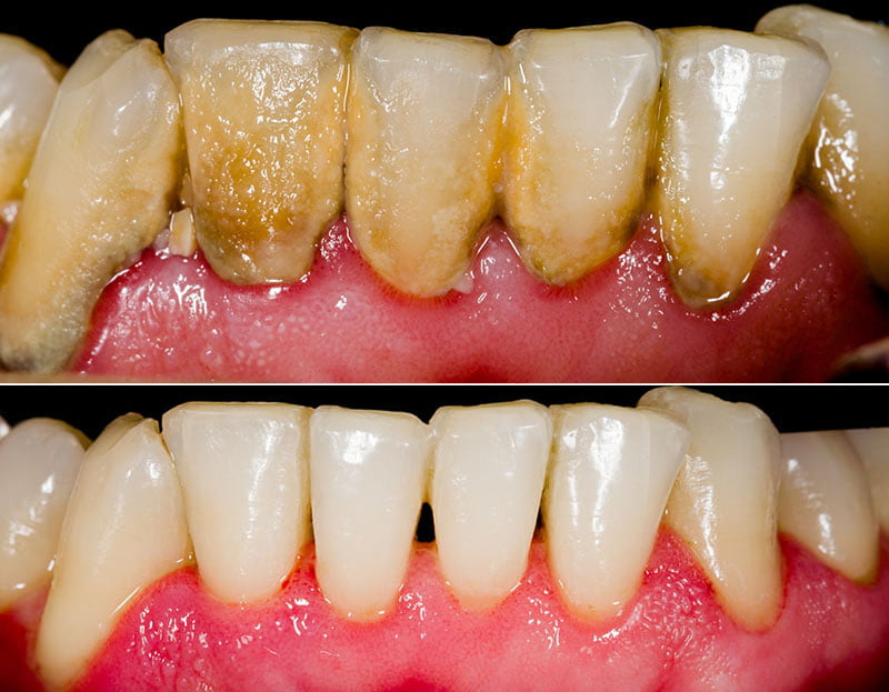
Gingivitis Gingivitis is a common and mild form of gum disease (periodontal disease) that causes irritation, redness and swelling (inflammation) of your gingiva, the part of your gum around the base of your teeth. It’s important to take gingivitis seriously and treat it promptly. Gingivitis can lead to much more serious gum disease called periodontitis and tooth loss. The most common cause of gingivitis is poor oral hygiene. Good oral health habits, such as brushing at least twice a day, flossing daily and getting regular dental checkups, can help prevent and reverse gingivitis. Causes of Gingivitis The most common cause of gingivitis is poor oral hygiene that encourages plaque to form on teeth, causing inflammation of the surrounding gum tissues. Here’s how plaque can lead to gingivitis: Plaque forms on your teeth.  Plaque is an invisible, sticky film composed mainly of bacteria that forms on your teeth when starches and sugars in food interact with bacteria normally found in your mouth. Plaque requires daily removal because it re-forms quickly. Plaque turns into tartar.  Plaque that stays on your teeth can harden under your gumline into tartar (calculus), which collects bacteria. Tartar makes plaque more difficult to remove, creates a protective shield for bacteria and causes irritation along the gumline. You need professional dental cleaning to remove tartar. Gingiva become inflamed (gingivitis).  The longer that plaque and tartar remain on your teeth, the more they irritate the gingiva, the part of your gum around the base of your teeth, causing inflammation. In time, your gums become  swollen and bleed easily. Tooth decay (dental caries) also may result. If not treated, gingivitis can advance to periodontitis and eventual tooth loss. Symptoms of Gingivitis Healthy gums are firm and pale pink and fitted tightly around the teeth. Signs and symptoms of gingivitis include: When to see a dentist? If you notice any signs and symptoms of gingivitis, schedule an appointment with your dentist. The sooner you seek care, the better your chances of reversing damage from gingivitis and preventing its progression to periodontitis. Risk factors Gingivitis is common, and anyone can develop it. Factors that can increase your risk of gingivitis include: pills Complications Untreated gingivitis can progress to gum disease that spreads to underlying tissue and bone (periodontitis), a much more serious condition that can lead to tooth loss. Chronic gingiva inflammation has been thought to be associated with some systemic diseases such as respiratory disease, diabetes, coronary artery disease, stroke and rheumatoid arthritis. Some research suggests that the bacteria responsible for periodontitis can enter your bloodstream through gum tissue, possibly affecting your heart, lungs and other parts of your body. But more studies are needed to confirm a link. Trench mouth, also known as necrotizing ulcerative gingivitis (NUG), is a severe form of gingivitis that causes painful, infected, bleeding gums and ulcerations. Trench mouth is rare today in developed nations, though it’s common in developing countries that have poor nutrition and poor living conditions. Prevention Good oral hygiene.  That means brushing your teeth for two minutes at least twice daily in the morning and before going to bed and flossing at least once a day. Better yet, brush after every meal or snack or as your dentist recommends. Flossing before you brush allows you to clean away the loosened food particles and bacteria. Regular dental visits.  See your dentist or dental hygienist regularly for cleanings, usually every six to 12 months. If you have risk factors that increase your chance of developing periodontitis — such as having dry mouth, taking certain medications or smoking — you may need professional cleaning more often. Annual dental X-rays can help identify diseases that are not seen by a visual dental examination and monitor for changes in your dental health. Good health practices.  Practices such as healthy eating and managing blood sugar if you have diabetes also are important to maintain gum health. Diagnosis Dentists usually diagnose gingivitis based on: Review of your dental and medical history and conditions that may contribute to your symptoms. Examination of your teeth, gums, mouth and tongue for signs of plaque and inflammation. Measuring the pocket depth of the groove between your gums and your teeth by inserting a dental probe beside your tooth beneath your gum line, usually at several sites throughout your mouth. In a healthy mouth, the pocket depth is usually between 1 and 3 millimeters (mm). Pockets deeper than 4 mm may indicate gum disease. Dental X-rays to check for bone loss in areas where your dentist sees deeper pockets. Other tests as needed.  If it’s not clear what has caused your gingivitis, your dentist may recommend that you get a medical evaluation to check for underlying health conditions. If your gum disease is advanced, your dentist may refer you to a specialist in gum diseases (periodontist). Treatment Prompt treatment usually reverses symptoms of gingivitis and prevents its progression to more serious gum disease and tooth loss. You have the best chance for successful treatment when you also adopt a daily routine of good oral care and stop tobacco use. Professional gingivitis care includes: Professional dental cleaning.  Your initial professional cleaning will include removing all traces of plaque, tartar and bacterial products a procedure known as scaling and root planing. Scaling removes tartar and bacteria from your tooth surfaces and beneath your gums. Root planning removes the bacterial products produced by inflammation, smooths the root surfaces, discouraging further buildup of tartar and bacteria, and allows proper healing. The procedure may be performed using instruments, a laser or an ultrasonic device. Dental restoration, if needed.  Misaligned teeth or poorly fitting crowns, bridges or other dental restorations may irritate your gums and make it harder to remove plaque during daily oral care. If problems with your teeth or dental restorations contribute to your gingivitis, your dentist may recommend fixing these problems. Ongoing care.  Gingivitis usually clears up after a thorough professional cleaning — as long as you continue good oral hygiene at home. Your dentist will

Gingivitis Read More »

Genital warts

Genital warts Genital warts are one of the most common types of sexually transmitted infections. Nearly all sexually active people will become infected with at least one type of human papillomavirus (HPV), the virus that causes genital warts, at some point during their lives. Women are somewhat more likely than men to develop genital warts. As the name suggests, genital warts affect the moist tissues of the genital area. Genital warts may look like small, flesh-colored bumps or have a cauliflower-like appearance. In many cases, the warts are too small to be visible. Like warts that appear elsewhere on your body, genital warts are caused by the human papillomavirus (HPV). Some strains of genital HPV can cause genital warts, while others can cause cancer. Vaccines can help protect against certain strains of genital HPV. Causes of Genital warts The human papillomavirus (HPV) causes warts. There are more than 40 different strains of HPV that specifically affect the genital area. Genital HPV is spread through sexual contact. In most cases, your immune system kills genital HPV and you never develop signs or symptoms of the infection. Symptoms of Genital warts In women, genital warts can grow on the vulva, the walls of the vagina, the area between the external genitals and the anus, the anal canal, and the cervix. In men, they may occur on the tip or shaft of the penis, the scrotum, or the anus. Genital warts can also develop in the mouth or throat of a person who has had oral sexual contact with an infected person. The signs and symptoms of genital warts include: N.B – Genital warts may be so small and flat that they can’t be seen with the naked eye. Sometimes, however, genital warts may multiply into large clusters. When to see a doctor? See a doctor if you or your partner develops bumps or warts in the genital area. Risk factors The Centers for Disease Control and Prevention estimates that nearly all sexually active people will become infected with at least one type of HPV at some point during their lives. Factors that can increase your risk of becoming infected include: Complications of Genital warts Genital wart complications may include: Cancer.  Cervical cancer has been closely linked with genital HPV infection. Certain types of HPV also are associated with cancer of the vulva, cancer of the anus, cancer of the penis, and cancer of the mouth and throat. Human papillomavirus infection doesn’t always lead to cancer, but it’s still important for women to have regular Pap tests, particularly if you’ve been infected with higher risk types of HPV. Problems during pregnancy. Genital  warts may cause problems during pregnancy. Warts could enlarge, making it difficult to urinate. Warts on the vaginal wall may reduce the ability of vaginal tissues to stretch during childbirth. Large warts on the vulva or in the vagina can bleed when stretched during delivery. Rarely, a baby born to a mother with genital warts may develop warts in his or her throat. The baby may need surgery to make sure his or her airway isn’t blocked. Prevention Using a condom every time you have sex can significantly reduce your risk of contracting genital warts. Although condom use can reduce your risk, it is not 100 percent effective. You can still get genital warts. Vaccination A vaccine known as Gardasil protects against four strains of HPV that cause cancer, and is used to prevent genital warts. In 2014, the U.S. Food and Drug Administration approved a vaccine called Gardasil 9, which protects against nine strains of HPV. Another vaccine, called Cervarix, protects against cervical cancer but not genital warts. The national Advisory Committee on Immunization Practices recommends routine HPV vaccination for girls and boys ages 11 and 12. If not fully vaccinated at that age, it’s recommended that girls and women through age 26 and boys and men through age 21 receive the vaccine. However, men may receive the HPV vaccine through age 26 if desired. These vaccines are most effective if given to children before they become sexually active. Research has shown that people under 21 and age 21 to 30 who receive the HPV vaccine can reduce their risk of getting genital warts by up to 50 percent. Side effects from the vaccines are usually mild and include soreness at the injection site (the upper arm), headaches, low-grade fever or flu-like symptoms. Sometimes dizziness or fainting occurs after the injection, especially in adolescents. Diagnosis of Genital warts Because it’s often difficult to detect genital warts, your doctor may apply a mild acetic acid solution to your genitals to whiten any warts. Then, he or she may view them through a special magnifying instrument, a colposcope. Pap tests For women, it’s important to have regular pelvic exams and Pap tests, which can help detect vaginal and cervical changes caused by genital warts or the early signs of cervical cancer a possible complication of genital HPV infection. During a Pap test, your doctor will use a device called a speculum to hold open your vagina. He or she will then use a long-handled tool to collect a small sample of cells from your cervix the passage between your vagina and your uterus. The cells are examined with a microscope for abnormalities. HPV test Only a few types of genital HPV have been linked to cervical cancer. A sample of cervical cells, taken during a Pap test, can be tested for these cancer-causing HPV strains. This test is generally reserved for women ages 30 and older. It isn’t as useful for younger women because their immune systems usually can kill even cancer-causing varieties of genital HPV without treatment. Treatment of Genital warts If your warts aren’t causing discomfort, you may not need treatment. But if your symptoms include itching, burning and pain, or if visible warts are causing emotional distress, your doctor can help you clear an outbreak with medications or surgery. However, the lesions are likely to come back after

Genital warts Read More »

Gastroesophageal reflux disease (GERD)

Gastroesophageal reflux disease (GERD) Gastroesophageal reflux disease (GERD) occurs when stomach acid frequently flows back into the tube connecting your mouth and stomach (esophagus). This backwash (acid reflux) can irritate the lining of your esophagus. Many people experience acid reflux from time to time. GERD is mild acid reflux that occurs at least twice a week, or moderate to severe acid reflux that occurs at least once a week. Most people can manage the discomfort of GERD with lifestyle changes and over-the-counter medications. But some people with GERD may need stronger medications or surgery to ease symptoms. Causes of Gastroesophageal reflux disease (GERD) GERD is caused by frequent acid reflux. When you swallow, a circular band of muscle around the bottom of your esophagus (lower esophageal sphincter) relaxes to allow food and liquid to flow into your stomach. Then the sphincter closes again. If the sphincter relaxes abnormally or weakens, stomach acid can flow back up into your esophagus. This constant backwash of acid irritates the lining of your esophagus, often causing it to become inflamed. Symptoms of Gastroesophageal reflux disease (GERD) Common signs and symptoms of GERD include: If you have nighttime acid reflux, you might also experience: When to see a doctor? Seek immediate medical care if you have chest pain, especially if you also have shortness of breath, or jaw or arm pain. These may be signs and symptoms of a heart attack. Make an appointment with your doctor if you: Experience severe or frequent GERD symptoms Take over-the-counter medications for heartburn more than twice a week Risk factors Conditions that can increase your risk of GERD include: Factors that can aggravate acid reflux include: Complications Over time, chronic inflammation in your esophagus can cause: Narrowing of the esophagus (esophageal stricture).  Damage to the lower esophagus from stomach acid causes scar tissue to form. The scar tissue narrows the food pathway, leading to problems with swallowing. An open sore in the esophagus (esophageal ulcer).  Stomach acid can wear away tissue in the esophagus, causing an open sore to form. An esophageal ulcer can bleed, cause pain and make swallowing difficult. Precancerous changes to the esophagus (Barrett’s esophagus).  Damage from acid can cause changes in the tissue lining the lower esophagus. These changes are associated with an increased risk of esophageal cancer. Diagnosis Your doctor might be able to diagnose GERD based on a physical examination and history of your signs and symptoms. To confirm a diagnosis of GERD, or to check for complications, your doctor might recommend: Upper endoscopy.  Your doctor inserts a thin, flexible tube equipped with a light and camera (endoscope) down your throat, to examine the inside of your esophagus and stomach. Test results can often be normal when reflux is present, but an endoscopy may detect inflammation of the esophagus (esophagitis) or other complications. An endoscopy can also be used to collect a sample of tissue (biopsy) to be tested for complications such as Barrett’s esophagus. Ambulatory acid (pH) probe test.  A monitor is placed in your esophagus to identify when, and for how long, stomach acid regurgitates there. The monitor connects to a small computer that you wear around your waist or with a strap over your shoulder. The monitor might be a thin, flexible tube (catheter) that’s threaded through your nose into your esophagus, or a clip that’s placed in your esophagus during an endoscopy and that gets passed into your stool after about two days. Esophageal manometry.  This test measures the rhythmic muscle contractions in your esophagus when you swallow. Esophageal manometry also measures the coordination and force exerted by the muscles of your esophagus. X-ray of your upper digestive system.  X-rays are taken after you drink a chalky liquid that coats and fills the inside lining of your digestive tract. The coating allows your doctor to see a silhouette of your esophagus, stomach and upper intestine. You may also be asked to swallow a barium pill that can help diagnose a narrowing of the esophagus that may interfere with swallowing. Treatment Your doctor is likely to recommend that you first try lifestyle modifications and over-the-counter medications. If you don’t experience relief within a few weeks, your doctor might recommend prescription medication or surgery. Over-the-counter medications The options include: Antacids that neutralize stomach acid.  Antacids, such as Mylanta, Rolaids and Tums, may provide quick relief. But antacids alone won’t heal an inflamed esophagus damaged by stomach acid. Overuse of some antacids can cause side effects, such as diarrhea or sometimes kidney problems. Medications to reduce acid production.  These medications  known as H-2-receptor blockers include cimetidine (Tagamet HB), famotidine (Pepcid AC), nizatidine (Axid AR) and ranitidine. H-2-receptor blockers don’t act as quickly as antacids, but they provide longer relief and may decrease acid production from the stomach for up to 12 hours. Stronger versions are available by prescription. Medications that block acid production and heal the esophagus.  These medications  known as proton pump inhibitors are stronger acid blockers than H-2-receptor blockers and allow time for damaged esophageal tissue to heal. Over-the-counter proton pump inhibitors include lansoprazole (Prevacid 24 HR) and omeprazole (Prilosec OTC, Zegerid OTC). Prescription medications Prescription-strength treatments for GERD include: Prescription-strength H-2-receptor blockers.  These include prescription-strength famotidine (Pepcid), nizatidine and ranitidine. These medications are generally well-tolerated but long-term use may be associated with a slight increase in risk of vitamin B-12 deficiency and bone fractures. Prescription-strength proton pump inhibitors.  These include esomeprazole (Nexium), lansoprazole (Prevacid), omeprazole (Prilosec, Zegerid), pantoprazole (Protonix), rabeprazole (Aciphex) and dexlansoprazole (Dexilant). Although generally well-tolerated, these medications might cause diarrhea, headache, nausea and vitamin B-12 deficiency. Chronic use might increase the risk of hip fracture. Medication to strengthen the lower esophageal sphincter.  Baclofen may ease GERD by decreasing the frequency of relaxations of the lower esophageal sphincter. Side effects might include fatigue or nausea. Surgery and other procedures GERD can usually be controlled with medication.  But if medications don’t help or you wish to avoid long-term medication use, your doctor might recommend: Fundoplication.  The surgeon

Gastroesophageal reflux disease (GERD) Read More »

Bedsores (pressure ulcers)

Bedsores (pressure ulcers) Bedsores also called pressure ulcers and decubitus ulcers are injuries to skin and underlying tissue resulting from prolonged pressure on the skin. Bedsores most often develop on skin that covers bony areas of the body, such as the heels, ankles, hips and tailbone. People most at risk of bedsores are those with a medical condition that limits their ability to change positions or those who spend most of their time in a bed or chair. Bedsores can develop quickly. Most sores heal with treatment, but some never heal completely. You can take steps to help prevent bedsores and aid healing. Causes of Bedsores Bedsores are caused by pressure against the skin that limits blood flow to the skin. Other factors related to limited mobility can make the skin vulnerable to damage and contribute to the development of pressure sores. Three primary contributing factors for bedsores are: Pressure.  Constant pressure on any part of your body can lessen the blood flow to tissues. Blood flow is essential to delivering oxygen and other nutrients to tissues. Without these essential nutrients, skin and nearby tissues are damaged and might eventually die. For people with limited mobility, this kind of pressure tends to happen in areas that aren’t wellpadded with muscle or fat and that lie over a bone, such as the spine, tailbone, shoulder blades, hips, heels and elbows. Friction.  Friction occurs when the skin rubs against clothing or bedding. It can make fragile skin more vulnerable to injury, especially if the skin is also moist. Shear.  Shear occurs when two surfaces move in the opposite direction. For example, when a bed is elevated at the head, you can slide down in bed. As the tailbone moves down, the skin over the bone might stay in place essentially pulling in the opposite direction. Symptoms of Bedsores Warning signs of pressure ulcers are: N.B – Bedsores fall into one of several stages based on their depth, severity and other characteristics. The degree of skin and tissue damage ranges from red, unbroken skin to a deep injury involving muscle and bone. Common sites of pressure sores For people who use a wheelchair, pressure sores often occur on skin over the following sites: For people who are confined to a bed, common sites include the following: When to see a doctor? If you notice warning signs of a bedsore, change your position to relieve the pressure on the area. If you don’t see improvement in 24 to 48 hours, contact your doctor. Seek immediate medical care if you show signs of infection, such as a fever, drainage from a sore, a sore that smells bad, or increased redness, warmth or swelling around a sore. Risk factors People are at risk of developing pressure sores if they have difficulty moving and are unable to easily change position while seated or in bed. Risk factors include: Immobility.  This might be due to poor health, spinal cord injury and other causes. Lack of sensory perception.  Spinal cord injuries, neurological disorders and other conditions can result in a loss of sensation. An inability to feel pain or discomfort can result in not being aware of warning signs and the need to change position. Poor nutrition and hydration.  People need enough fluids, calories, protein, vitamins and minerals in their daily diet to maintain healthy skin and prevent the breakdown of tissues. Medical conditions affecting blood flow.  Health problems that can affect blood flow, such as diabetes and vascular disease, increase the risk of tissue damage. Complications Complications of pressure ulcers, some life-threatening, include: Cellulitis.  Cellulitis is an infection of the skin and connected soft tissues. It can cause warmth, redness and swelling of the affected area. People with nerve damage often do not feel pain in the area affected by cellulitis. Bone and joint infections.  An infection from a pressure sore can burrow into joints and bones. Joint infections (septic arthritis) can damage cartilage and tissue. Bone infections (osteomyelitis) can reduce the function of joints and limbs. Cancer.  Long-term, nonhealing wounds (Marjolin’s ulcers) can develop into a type of squamous cell carcinoma Sepsis.  Rarely, a skin ulcer leads to sepsis. Prevention You can help prevent bedsores by frequently repositioning yourself to avoid stress on the skin. Other strategies include taking good care of your skin, maintaining good nutrition and fluid intake, quitting smoking, managing stress, and exercising daily. Tips for repositioning Consider the following recommendations related to repositioning in a bed or chair: Shift your weight frequently.  If you use a wheelchair, try shifting your weight about every 15 minutes. Ask for help with repositioning about once an hour. Lift yourself, if possible.  If you have enough upper body strength, do wheelchair pushups raising your body off the seat by pushing on the arms of the chair. Look into a specialty wheelchair.  Some wheelchairs allow you to tilt them, which can relieve pressure. Select cushions or a mattress that relieves pressure.  Use cushions or a special mattress to relieve pressure and help ensure your body is wellpositioned. Do not use doughnut cushions, as they can focus pressure on surrounding tissue. Adjust the elevation of your bed.  If your bed can be elevated at the head, raise it no more than 30 degrees. This helps prevent shearing. Tips for skin care Consider the following suggestions for skin care: Keep skin clean and dry.  Wash the skin with a gentle cleanser and pat dry. Do this cleansing routine regularly to limit the skin’s exposure to moisture, urine and stool. Protect the skin.  Use plain talcum powder to protect skin at friction points. Apply lotion to dry skin. Change bedding and clothing frequently if needed. Watch for buttons on the clothing and wrinkles in the bedding that irritate the skin. Inspect the skin daily.  Look closely at your skin daily for warning signs of a pressure sore. Diagnosis Your doctor will look closely at your skin to determine whether you have a pressure ulcer and how bad the damage is.

Bedsores (pressure ulcers) Read More »

Trichomoniasis

 Trichomoniasis Trichomoniasis is a common sexually transmitted infection caused by a parasite. In women, trichomoniasis can cause a foul-smelling vaginal discharge, genital itching and painful urination. Men who have trichomoniasis typically have no symptoms. Pregnant women who have trichomoniasis might be at higher risk of delivering their babies prematurely. To prevent reinfection with the organism that causes trichomoniasis, both partners should be treated. The most common treatment for trichomoniasis involves taking one megadose of metronidazole (Flagyl) or tinidazole (Tindamax). You can reduce your risk of infection by using condoms correctly every time you have sex. Causes of Trichomoniasis Trichomoniasis is caused by a one-celled protozoan, a type of tiny parasite that travels between people during sexual intercourse. The incubation period between exposure and infection is unknown, but it’s thought to range from five to 28 days. Symptoms of  Trichomoniasis Many women and most men with trichomoniasis have no symptoms, at least not at first. Trichomoniasis signs and symptoms for women include: N.B -Trichomoniasis rarely causes symptoms in men.  When men do have signs and symptoms, however, they might include: When to see a doctor See your doctor if you have a foul-smelling vaginal discharge or if you have pain with urination or sexual intercourse. Risk factors Risk factors include having: Complications Pregnant women who have trichomoniasis might:  Prevention As with other sexually transmitted infections, the only way to prevent trichomoniasis is to abstain from sex. To lower your risk, use condoms correctly every time you have sex. Diagnosis The diagnosis of trichomoniasis can be confirmed by looking at a sample of vaginal fluid for women or urine for men under a microscope. Growing a culture used to be the way to diagnose trichomoniasis, but newer, faster tests, such as rapid antigen tests and nucleic acid amplification, are more common now. Treatment The most common treatment for trichomoniasis, even for pregnant women, is to swallow one megadose of either metronidazole (Flagyl) or tinidazole (Tindamax). In some cases, your doctor might recommend a lower dose of metronidazole two times a day for seven days. Both you and your partner need treatment. And you need to avoid sexual intercourse until the infection is cured, which takes about a week. Don’t drink alcohol for 24 hours after taking metronidazole or 72 hours after taking tinidazole, because it can cause severe nausea and vomiting. Your doctor will likely want to retest you for trichomoniasis from two weeks to three months after treatment to be sure you haven’t been reinfected. N.B – Untreated, trichomoniasis can last for months to years. Visit us on : www.healthalert.co.za    Calls us on : +27 82 0941 375   Email us on : info@healthalert.co.za  

Trichomoniasis Read More »

Cataracts

Cataracts A cataract is a clouding of the normally clear lens of your eye. For people who have cataracts, seeing through cloudy lenses is a bit like looking through a frosty or fogged-up window. Clouded vision caused by cataracts can make it more difficult to read, drive a car (especially at night) or see the expression on a friend’s face. Most cataracts develop slowly and don’t disturb your eyesight early on. But with time, cataracts will eventually interfere with your vision. At first, stronger lighting and eyeglasses can help you deal with cataracts. But if impaired vision interferes with your usual activities, you might need cataract surgery. Fortunately, cataract surgery is generally a safe, effective procedure. Causes of cataract Most cataracts develop when aging or injury changes the tissue that makes up your eye’s lens. Some inherited genetic disorders that cause other health problems can increase your risk of cataracts. Cataracts can also be caused by other eye conditions, past eye surgery or medical conditions such as diabetes. Long-term use of steroid medications, too, can cause cataracts to develop. Symptoms of cataract Signs and symptoms of cataracts include: N.B – At first, the cloudiness in your vision caused by a cataract may affect only a small part of the eye’s lens and you may be unaware of any vision loss. As the cataract grows larger, it clouds more of your lens and distorts the light passing through the lens. This may lead to more noticeable symptoms. When to see a doctor? Make an appointment for an eye exam if you notice any changes in your vision. If you develop sudden vision changes, such as double vision or flashes of light, sudden eye pain, or sudden headache, see your doctor right away. How a cataract forms The lens, where cataracts form, is positioned behind the colored part of your eye (iris). The lens focuses light that passes into your eye, producing clear, sharp images on the retina  the lightsensitive membrane in the eye that functions like the film in a camera. As you age, the lenses in your eyes become less flexible, less transparent and thicker. Age-related and other medical conditions cause tissues within the lens to break down and clump together, clouding small areas within the lens. As the cataract continues to develop, the clouding becomes denser and involves a bigger part of the lens. A cataract scatters and blocks the light as it passes through the lens, preventing a sharply defined image from reaching your retina. As a result, your vision becomes blurred. Cataracts generally develop in both eyes, but not evenly. The cataract in one eye may be more advanced than the other, causing a difference in vision between eyes. Types of cataracts Cataract types include: Cataracts affecting the center of the lens (nuclear cataracts).  A nuclear cataract may at first cause more nearsightedness or even a temporary improvement in your reading vision. But with time, the lens gradually turns more densely yellow and further clouds your vision. As the cataract slowly progresses, the lens may even turn brown.  Advanced yellowing or browning of the lens can lead to difficulty distinguishing between shades of color. Cataracts that affect the edges of the lens (cortical cataracts).  A cortical cataract begins as whitish, wedge-shaped opacities or streaks on the outer edge of the lens cortex. As it slowly progresses, the streaks extend to the center and interfere with light passing through the center of the lens. Cataracts that affect the back of the lens (posterior subcapsular cataracts).  A posterior subcapsular cataract starts as a small, opaque area that usually forms near the back of the lens, right in the path of light. A posterior subcapsular cataract often interferes with your reading vision, reduces your vision in bright light, and causes glare or halos around lights at night. These types of cataracts tend to progress faster than other types do. Cataracts you’re born with (congenital cataracts).  Some people are born with cataracts or develop them during childhood. These cataracts may be genetic, or associated with an intrauterine infection or trauma. These cataracts also may be due to certain conditions, such as myotonic dystrophy, galactosemia, neurofibromatosis type 2 or rubella. Congenital cataracts don’t always affect vision, but if they do they’re usually removed soon after detection. Risk factors Factors that increase your risk of cataracts include: Prevention No studies have proved how to prevent cataracts or slow the progression of cataracts. But doctors think several strategies may be helpful, including: Have regular eye examinations.  Eye examinations can help detect cataracts and other eye problems at their earliest stages. Ask your doctor how often you should have an eye examination. Quit smoking.  Ask your doctor for suggestions about how to stop smoking. Medications, counseling and other strategies are available to help you. Manage other health problems.  Follow your treatment plan if you have diabetes or other medical conditions that can increase your risk of cataracts. Choose a healthy diet that includes plenty of fruits and vegetables.  Adding a variety of colorful fruits and vegetables to your diet ensures that you’re getting many vitamins and nutrients. Fruits and vegetables have many antioxidants, which help maintain the health of your eyes. Studies haven’t proved that antioxidants in pill form can prevent cataracts.  But, a large population study recently showed that a healthy diet rich in vitamins and minerals was associated with a reduced risk of developing cataracts. Fruits and vegetables have many proven health benefits and are a safe way to increase the amount of minerals and vitamins in your diet. Wear sunglasses.  Ultraviolet light from the sun may contribute to the development of cataracts. Wear sunglasses that block ultraviolet B (UVB) rays when you’re outdoors. Reduce alcohol use.  Excessive alcohol use can increase the risk of cataracts. Diagnosis To determine whether you have a cataract, your doctor will review your medical history and symptoms, and perform an eye examination. Your doctor may conduct several tests, including: Visual acuity test.  A visual acuity test

Cataracts Read More »

Erectile dysfunction

Erectile dysfunction Erectile dysfunction (impotence) is the inability to get and keep an erection firm enough for sex. Having erection trouble from time to time isn’t necessarily a cause for concern. If erectile dysfunction is an ongoing issue, however, it can cause stress, affect your self-confidence and contribute to relationship problems. Problems getting or keeping an erection can also be a sign of an underlying health condition that needs treatment and a risk factor for heart disease. If you’re concerned about erectile dysfunction, talk to your doctor even if you’re embarrassed. Sometimes, treating an underlying condition is enough to reverse erectile dysfunction. In other cases, medications or other direct treatments might be needed. Causes of Erectile dysfunction Male sexual arousal is a complex process that involves the brain, hormones, emotions, nerves, muscles and blood vessels. Erectile dysfunction can result from a problem with any of these. Likewise, stress and mental health concerns can cause or worsen erectile dysfunction. Sometimes a combination of physical and psychological issues causes erectile dysfunction. For instance, a minor physical condition that slows your sexual response might cause anxiety about maintaining an erection. The resulting anxiety can lead to or worsen erectile dysfunction. Physical causes of erectile dysfunction In many cases, erectile dysfunction is caused by something physical. Common causes include: The brain plays a key role in triggering the series of physical events that cause an erection, starting with feelings of sexual excitement. A number of things can interfere with sexual feelings and cause or worsen erectile dysfunction. These include: Symptoms of Erectile dysfunction Erectile dysfunction symptoms might include persistent: When to see a doctor? A family doctor is a good place to start when you have erectile problems. See your doctor if: Risk factors As you get older, erections might take longer to develop and might not be as firm. You might need more direct touch to your penis to get and keep an erection. Various risk factors can contribute to erectile dysfunction, including: Complications Complications resulting from erectile dysfunction can include: Prevention The best way to prevent erectile dysfunction is to make healthy lifestyle choices and to manage any existing health conditions. For example: Diagnosis For many men, a physical exam and answering questions (medical history) are all that’s needed for a doctor to diagnose erectile dysfunction and recommend a treatment. If you have chronic health conditions or your doctor suspects that an underlying condition might be involved, you might need further tests or a consultation with a specialist. Tests for underlying conditions might include: Physical exam.  This might include careful examination of your penis and testicles and checking your nerves for sensation. Blood tests. A sample of your blood might be sent to a lab to check for signs of heart disease, diabetes, low testosterone levels and other health conditions. Urine tests (urinalysis). Like blood tests, urine tests are used to look for signs of diabetes and other underlying health conditions. Ultrasound. This test is usually performed by a specialist in an office. It involves using a wandlike device (transducer) held over the blood vessels that supply the penis. It creates a video image to let your doctor see if you have blood flow problems. This test is sometimes done in combination with an injection of medications into the penis to stimulate blood flow and produce an erection. Psychological exam. Your doctor might ask questions to screen for depression and other possible psychological causes of erectile dysfunction. Treatment The first thing your doctor will do is to make sure you’re getting the right treatment for any health conditions that could be causing or worsening your erectile dysfunction. Depending on the cause and severity of your erectile dysfunction and any underlying health conditions, you might have various treatment options. Your doctor can explain the risks and benefits of each treatment and will consider your preferences. Your partner’s preferences also might play a role in your treatment choices. Oral medications Oral medications are a successful erectile dysfunction treatment for many men. They include: N.B – All four medications enhance the effects of nitric oxide a natural chemical your body produces that relaxes muscles in the penis. This increases blood flow and allows you to get an erection in response to sexual stimulation. Taking one of these tablets will not automatically produce an erection. Sexual stimulation is needed first to cause the release of nitric oxide from your penile nerves. These medications amplify that signal, allowing some men to function normally. Oral erectile dysfunction medications are not aphrodisiacs, will not cause excitement and are not needed in men who get normal erections. The medications vary in dosage, how long they work and side effects. Possible side effects include flushing, nasal congestion, headache, visual changes, backache and stomach upset. Your doctor will consider your particular situation to determine which medication might work best. These medications might not treat your erectile dysfunction immediately. You might need to work with your doctor to find the right medication and dosage for you. Before taking any medication for erectile dysfunction, including over-the-counter supplements and herbal remedies, get your doctor’s OK. Medications for erectile dysfunction do not work in all men and might be less effective in certain conditions, such as after prostate surgery or if you have diabetes. Some medications might also be dangerous if you: Other medications Other medications for erectile dysfunction include: Alprostadil self-injection. With this method, you use a fine needle to inject alprostadil (Caverject Impulse, Edex) into the base or side of your penis. In some cases, medications generally used for other conditions are used for penile injections on their own or in combination. Examples include papaverine, alprostadil and phentolamine. Often these combination medications are known as bimix (if two medications are included) or trimix (if three are included). Each injection is dosed to create an erection lasting no longer than an hour. Because the needle used is very fine, pain from the injection site is usually minor. Side effects can include mild bleeding from

Erectile dysfunction Read More »